Running Archives - Opus Biological
Importance of calf capacity

Importance of calf capacity

The Importance of Good Calf Muscle Capacity in Preventing and Rehabilitating Lower Limb Injuries

Calf muscles, primarily consisting of the gastrocnemius and soleus muscles, play a vital role in lower limb function and overall biomechanics. These muscles are essential for movements such as walking, running, and jumping, contributing to ankle stability, propulsion, and shock absorption. Strong and flexible calf muscles not only enhance athletic performance but also prevent lower limb injuries and support rehabilitation after injury. This article outlines the importance of calf muscle capacity, provides data on its impact on joint loading, and explains how to assess calf capacity in clinical settings.

The Role of Calf Muscles in Lower Limb Function

The calf muscles work as a dynamic powerhouse during movement. The gastrocnemius and soleus muscles, in conjunction with the Achilles tendon, drive plantarflexion, which is the action of pushing the foot off the ground. This movement is crucial for walking, running, and jumping, as it propels the body forward. Additionally, the calf muscles play a role in absorbing the impact forces generated when the foot strikes the ground, thus reducing stress on other structures like the knee, hip, and lower back.

By acting as shock absorbers and stabilizers, the calf muscles influence the load distribution across the entire lower limb. Proper calf function ensures that forces are dispersed evenly, preventing overloading of other structures and contributing to balanced biomechanics. When calf muscles are weak or fatigued, they fail to perform this role efficiently, leading to improper movement patterns and potentially contributing to injuries of the lower limb, such as ankle sprains, Achilles tendinopathy, anterior knee pain, and plantar fasciitis. Weak calf musculature may also exacerbate symptoms such as osteoarthritis in the foot, ankle and knee.  A study by Alshami and Alhassany (2020) 1, found that calf muscle strength was significantly reduced in the patients with knee OA compared with that in the healthy control participants. Therefore it could be a good idea to ensure that within an OA knee patient’s exercise programme, there is targeted calf strengthening.

Impact of Strong Calf Muscles on Knee Joint Loading

Strong calf muscles not only support the ankle but also significantly reduce the load on the knee joint during various activities. Research has shown that adequate calf strength can reduce knee joint loading by absorbing the ground reaction forces that occur during walking or running. The soleus muscle, in particular, is known to play a key role in reducing the stress on the knee by generating posterior shear forces at the tibia, which counteract the forward forces that tend to overwork the knee.

A 2019 study by A. Bohm, 2,  found that the soleus muscle can contribute up to 50% of the force needed to decelerate the body during walking and running, thereby offloading the knee joint considerably. Research indicates that strong calf muscles can reduce the load on the knee by up to 20-25% during activities like walking and stair climbing . This effect is critical in both preventing injuries such as patellofemoral pain syndrome and in managing conditions like osteoarthritis, where reduced joint loading can slow the progression of cartilage wear.

In individuals with weakened calf muscles, this offloading effect is diminished, forcing the knee and other structures to take on additional load, which increases the risk of injury. Athletes, in particular, benefit from strong calf muscles, as these muscles allow them to handle high-impact activities like running and jumping without overstressing the knee joint.

Testing Calf Capacity in Clinical Settings

Given the importance of calf muscle capacity in preventing and rehabilitating lower limb injuries, clinicians need reliable methods to assess both the strength and endurance of the calf muscles. Below are some of the most effective ways to test calf capacity in a clinical setting:

  • Calf-Raise Test (Repetitive Heel-Raise Test): The single-leg calf-raise test is a simple and widely used method to assess calf muscle endurance. In this test, the patient stands on one leg and performs heel raises, lifting the heel off the ground as high as possible with the number of repetitions completed before fatigue, recorded. This test evaluates both the gastrocnemius and soleus muscles, providing a measure of endurance, which is critical for injury prevention. Hebert et. Al (2017) 2, carried out a great study which generated a guide to target rep ranges for different patient groups based on age and sex.
  • Maximal Voluntary Isometric Contraction (MVIC): This test measures the maximum force a person can generate during an isometric contraction of the calf muscles. Using a dynamometer, clinicians can measure the force output when the patient pushes against resistance in a fixed position. The MVIC is a reliable method to assess the maximal strength of the calf muscles, especially useful in identifying deficits that may contribute to injury risk or delay rehabilitation.
  • Single-Leg Hop Test: This test is used to evaluate functional strength and power in the calf muscles. The patient is asked to perform a series of single-leg hops for distance or height. A significant discrepancy between the two legs indicates potential calf muscle weakness or imbalance. This test is particularly useful for athletes who need to perform explosive movements during sport, as it mimics the high-force demands placed on the calf muscles.
  • Functional Movement Assessments: Assessing how the calf muscles function during movements like squats, walking, or running is also valuable in clinical settings. Analysing gait or performing functional movement screens allows clinicians to observe whether the calf muscles are functioning optimally during dynamic activities. This type of assessment is particularly relevant for athletes and individuals recovering from injury, as it highlights any biomechanical deficiencies related to calf muscle performance.

Rehabilitation of Lower Limb Injuries: Calf Strengthening

Injury rehabilitation programs often include exercises aimed at restoring calf muscle strength, endurance, and flexibility. Isometric calf exercises can be used early in rehabilitation to improve muscle activation without placing too much strain on the injured tissue. Over time, patients progress to more dynamic exercises such as calf raises, hopping, and plyometric drills, depending on the severity of their injury and their physical goals. Green et. Al (2022) 3, break up the involvement of calf specific exercises into 4 nice stages which a patient or player should go through in order to go from day 1 to return to play. These stages are: Early loading and foundation calf and lower limb function, Loaded strengthening, loaded power, plyometrics and ballistics and finally, Locomotion.

Conclusion

Good calf muscle capacity is vital for preventing lower limb injuries and supporting effective rehabilitation. Strong and flexible calf muscles reduce the load on the knee joint by as much as 20-25%, play an essential role in stabilizing the ankle, and help prevent common injuries such as Achilles tendinopathy, plantar fasciitis, and knee injuries. Clinicians can use a variety of tests, such as the calf-raise test and MVIC, to assess calf muscle strength and endurance. By incorporating calf training and flexibility exercises into rehabilitation and prevention programs, individuals can improve their lower limb function, reduce the risk of injury, and ensure a quicker recovery following injury.

Reference List


  • Alshami, A.M. and Alhassany, H.A. (2020). Girth, strength, and flexibility of the calf muscle in patients with knee osteoarthritis: A case–control study. Journal of Taibah University Medical Sciences, 15(3), pp.197–202. doi:https://doi.org/10.1016/j.jtumed.2020.04.002.
  • Bohm S, Mersmann F, Santuz A, Arampatzis A.. 2019. The force–length–velocity potential of the human soleus muscle is related to the energetic cost of running. Proc. R. Soc. B Biol. Sci. 286, 20192560 ( 10.1098/rspb.2019.2560)
  • Hébert-Losier, K., Wessman, C., Alricsson, M. and Svantesson, U. (2017). Updated reliability and normative values for the standing heel-rise test in healthy adults. Physiotherapy, 103(4), pp.446–452. doi:https://doi.org/10.1016/j.physio.2017.03.002.
  • Green, B., McClelland, J.A., Semciw, A.I. et al. The Assessment, Management and Prevention of Calf Muscle Strain Injuries: A Qualitative Study of the Practices and Perspectives of 20 Expert Sports Clinicians. Sports Med – Open 8, 10 (2022). https://doi.org/10.1186/s40798-021-00364-0
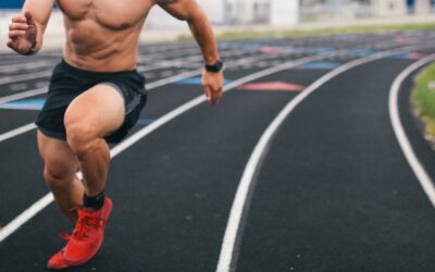
Runner in shorts and trainers on an outdoor athletics track
Sometimes – less means more

Sometimes – less means more

Unfortunately, sometimes we come face to face with patients who have a rather long road ahead of them when it comes to returning to their pre-injury status. An ACL or Achilles reconstruction for example. This means a longer period for them to be vigilant, compliant and motivated with their session attendance, exercise and management techniques in and away from face-to-face sessions. I for one know that if I was placed in this scenario, I would find it tough to keep the same level of determination to adhere to my programme from beginning to end. So, can we use a little something to aid this lengthy process for our patients which will not negatively impact their progress? In my opinion, yes. Deloading.

Bell et. al (2023) (1) define ‘Deloading’ as a period of reduced training stress designed to mitigate physiological and psychological fatigue, promote recovery, and enhance preparedness for subsequent training.

In the realm of sports rehabilitation, the concept of deload periods has gained significant traction as an approach to managing and enhancing recovery from injuries. Deload periods, often implemented within a structured rehabilitation program, involve a planned reduction in exercise intensity and or volume. This technique is particularly beneficial in long-term recovery processes, offering numerous physiological and psychological benefits that can expedite healing and improve overall outcomes

The primary physiological benefit of deload periods is the mitigation of overtraining and excessive fatigue (Rogerson et.al 2024) (5). During a prolonged rehabilitation process, continuous high-intensity training can lead to increased stress on the injured tissues, potentially exacerbating the injury or slowing down the healing process. By incorporating deload periods, the body is afforded the necessary time to recover and adapt to the rehabilitation exercises without being overwhelmed by continuous strain.

Deload periods can also play a crucial role in preventing the risk of re-injury. As targeted structures are gradually strengthened during rehabilitation, they require adequate rest to fully recover and adapt to the increased loads. Without sufficient recovery, these structures remain vulnerable to further damage. As per Mosewich, Kent and Kowalski (2013) (4) deloading helps ensure that the healing tissues are not subjected to undue stress, thus reducing the likelihood of setbacks.

Additionally, deload periods facilitate metabolic recovery. Intense exercise sessions can deplete glycogen stores, disrupt hormonal balances, and lead to an accumulation of metabolic byproducts. A period of reduced training intensity allows for the replenishment of glycogen stores, normalization of hormone levels, and clearance of metabolic waste, thereby optimizing the body’s readiness for subsequent training phases (Ivy 2004) (3).

Beyond the physiological advantages, deload periods offer substantial psychological benefits that are vital for a successful rehabilitation journey. Long-term injury recovery can be mentally taxing, often leading to feelings of frustration, anxiety, or even burnout. Scheduled deload periods provide individuals with a break, helping to alleviate mental fatigue and maintain motivation throughout the rehabilitation process. It is a well-known fact that within elite sport it is common for a player to be advised to go away to somewhere warm to put their feet up for 7-10 days at a certain stage of their recovery. This gets the player away from the current rehabilitation setting, enabling them to switch off mentally and relax.

The psychological relief afforded by deload periods also promotes adherence to the rehabilitation program (Bell et. al 2022) (2). Consistently high levels of training intensity can lead to a sense of dread or reluctance towards rehabilitation sessions. By incorporating periodic reductions in training demands, individuals are more likely to remain engaged and committed to their recovery plan, ultimately leading to better long-term outcomes

The implementation of deload periods within a rehabilitation program should be tailored to the individual’s specific injury, recovery progress, and overall training load. Generally, deload periods are scheduled every 6-8 weeks, but this can vary based on the intensity and frequency of the rehabilitation exercises. Monitoring the patient’s feedback, progress, and any signs of overtraining or fatigue can help in deciding the optimum time for this process also.  During a deload week, exercise intensity and volume are typically reduced by 50-70%, allowing the body ample time to recover without completely halting progress.

So, I think based on the above, it is safe to say that as therapists we should very much consider the implementation of the deload principle into the rehabilitation plans of those patients who have a longer and slightly more mentally and physically testing battle ahead. It is a big part of our role to support our patient’s and to keep them on the right track. Sometimes that may mean periodically seeing them and doing less to achieve more down the line.

Reference List


  • Bell, L., Ben William Strafford, Coleman, M., Patroklos Androulakis-Korakakis and Nolan, D. (2023). Integrating Deloading into Strength and Physique Sports Training Programmes: An International Delphi Consensus Approach. Sports Medicine – Open, 9(1). doi:https://doi.org/10.1186/s40798-023-00633-0.
  • Bell, L., Nolan, D., Immonen, V., Helms, E., Dallamore, J., Wolf, M. and Androulakis Korakakis, P. (2022). ‘You can’t shoot another bullet until you’ve reloaded the gun’: Coaches’ perceptions, practices and experiences of deloading in strength and physique sports. Frontiers in Sports and Active Living, [online] 4. doi:https://doi.org/10.3389/fspor.2022.1073223.
  • Ivy JL. Regulation of muscle glycogen repletion, muscle protein synthesis and repair following exercise. J Sports Sci Med. 2004 Sep 1;3(3):131-8. PMID: 24482590; PMCID: PMC3905295.
  • Amber D. Mosewich , Peter R.E. Crocker & Kent C. Kowalski (2013): Managinginjury and other setbacks in sport: experiences of (and resources for) high-performance women athletes, Qualitative Research in Sport, Exercise and Health, DOI:10.1080/2159676X.2013.766810
  • Rogerson, D., Nolan, D., Korakakis, P.A. et al. Deloading Practices in Strength and Physique Sports: A Cross-sectional Survey. Sports Med – Open 10, 26 (2024). https://doi.org/10.1186/s40798-024-00691-y
Person resting in bed, shot in black and white
Recovering from Delayed Onset Muscle Soreness

Recovering from Delayed Onset Muscle Soreness

Delayed onset muscle soreness (DOMS) is defined as ‘ultrastructural damage of muscle cells due to unfamiliar sporting activities or eccentric exercise, which leads to further protein degradation, apoptosis and local inflammatory response’ (Hotfiel et. al 2018) The micro tears caused by eccentric movements can impact performance by reducing joint range of motion and alter muscle recruitment patterns. This can increase risk of a soft tissue injury but treatment strategies for DOMS remain uncertain. Common treatment strategies include anti-inflammatories, massage and cryotherapy. Seidel et. al investigated the optimum treatment for DOMS and found non-steroidal anti-inflammatory drugs did reduce the pain but delayed the recovery.  Other interventions were examined (nutritional and physical) and found that there was a reduction in inflammation but no treatment aided muscle regeneration. (Seidel et. al, 2012)

Massage can be an effective tool to aid recovery from DOMS, however most research states the type and timing of the massage is important.  Hilbert et. al found that there was a reduction in muscle soreness 48 hours post exercise when massage is administered 2 hours after exercise, (Hilbert et. al, 2003). However, Visconti et. al found massage to be an effective tool to reduce DOMS during the onset of symptoms (Visconti et. al, 2015).

Cryotherapy has conflicting research on the effectiveness to alleviate DOMS. For example, Eston and Peters studied the use of cold-water immersion for recovery. They found 2-3 days post exercise, participants who used cold water immersion had increased joint range and reduced creatine kinase activity compared to the control group. However, both groups presented with muscle tenderness, swelling and reduced isometric strength 3 days following exercise. Howaston and Van Someren investigated the impact of ice massage therapy on DOMs, however discovered it is not an effective treatment due to only noticing creatine kinase reduction at 72 hours (Bishop et. al, 2008).

Evidence suggests adapting your exercise programme is the most efficient method to alleviate DOMS however the analgesic effect has been shown to be temporary (Zainuddin et. al, 2011). Cheung et. al suggests when exercising on a daily basis, one should reduce intensity and duration of exercise 1-2 days following DOMS. Training body parts that are less affected by DOMS and progressively overloading eccentric exercises over a 1 to 2 week period are efficient methods to manage DOMS (Cheung et. al 2003).

Reference List

Bishop, P.A., Jones, E. and Woods, A.K. (2008). Recovery From Training: A Brief Review. Journal of Strength and Conditioning Research, [online] 22(3), pp.1015–1024. doi:https://doi.org/10.1519/jsc.0b013e31816eb518.

Cheung, K., Hume, P.A. and Maxwell, L. (2012). Delayed Onset Muscle Soreness. Sports Medicine, [online] 33(2), pp.145–164. doi:https://doi.org/10.2165/00007256-200333020-00005.

Hilbert, J.E., Sforzo, G.A. and Swensen, T. (2003). The effects of massage on delayed onset muscle soreness. British Journal of Sports Medicine, [online] 37(1), pp.72–75. doi:https://doi.org/10.1136/bjsm.37.1.72.

Hotfiel, T., Freiwald, J., Hoppe, M., Lutter, C., Forst, R., Grim, C., Bloch, W., Hüttel, M. and Heiss, R. (2018). Advances in Delayed-Onset Muscle Soreness (DOMS): Part I: Pathogenesis and Diagnostics. Sportverletzung · Sportschaden, 32(04), pp.243–250. doi:https://doi.org/10.1055/a-0753-1884.

Seidel, E., Rother, M., Hartmann, J., Rother, I., Schaaf, T., Winzer, M., Fischer, A. and Regenspurger, K. (2012). Eccentric Exercise and Delayed Onset of Muscle Soreness (DOMS) – an Overview. Physikalische Medizin, Rehabilitation Medizin, Kurortmedizin, 22(02), pp.57–63. doi:https://doi.org/10.1055/s-0032-1304576.

Visconti, L., Capra, G., Carta, G., Forni, C. and Janin, D. (2015). Effect of massage on DOMS in ultramarathon runners: A pilot study. Journal of Bodywork and Movement Therapies, [online] 19(3), pp.458–463. doi:https://doi.org/10.1016/j.jbmt.2014.11.008.

Zainuddin, Z., Sacco, P., Newton, M. and Nosaka, K. (2006). Light concentric exercise has a temporarily analgesic effect on delayed-onset muscle soreness, but no effect on recovery from eccentric exercise. Applied Physiology, Nutrition, and Metabolism, 31(2), pp.126–134. doi:https://doi.org/10.1139/h05-010.

Man raising his arm behind his head while his shoulder is assessed
Why are injuries so common in athletes?

Why are injuries so common in athletes?

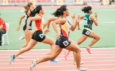
Group of women sprinting on an outdoor athletics track
Photo by Jonathan Chng on Unsplash

Sports injuries are extremely common, with figures estimating that amongst the younger population alone, over 3.5 million children and teens are injured each year as part of organised sports or physical activity. This does not even account for unreported injuries. Given their prevalence, it is important that we understand why injuries are more common amongst athletes and those that regularly participate in sporting activities so that we can prevent and treat these accordingly.

A sports injury can occur as a result of several scenarios, including accidents, impact, insufficient training, improper equipment, lack of conditioning, or poor warming up and stretching, to name a few.
Although joint injuries represent the most common site of damage, accounting for both sprains and strains, any part of your body can get injured during physical activity. When we exercise, a significant number of our muscles and tendons are involved; therefore, the chances of developing an injury are much greater.
Moreover, especially during competitive sporting activities, we tend to stretch our bodies and push ourselves farther than we usually would, resulting in us moving in ways that we often wouldn’t. Although this can be harmless, in some instances, this leaves our muscles and tendons torn, damaged, and in pain.

In athletes, the above factors are exacerbated, with the most common sports injuries amongst this population being:

  1. Strains: A strain is defined as an injury to either a muscle or a tendon that primarily results from overuse, force, or stretching. The severity of the injury depends on the nature of its cause, with strains ranging from a simple overstretching of the muscle or tendon to a partial or complete tear.
  2. Sprains: Alongside strains, sprains represent the most common type of injury across all sports, being especially common amongst athletes. A sprain is characterised by a torn muscle or ligament that results from overuse or excessive force, whether that be a collision, a fall, or other causes.
  3. Knee injuries: Damage to the knee joint is most common in contact sports and is usually caused by a sprain, strain or tendonitis. There are a vast number of different conditions that may result from an injury to the knee, including ACL tears, MCL/LCL ruptures, runner’s knee, IT band syndrome and jumper’s knee, anterior knee pain, and patella-femoral maltracking.
  4. Fractures: The most prevalent fractures seen amongst athletes are fractured wrists, hands, collarbones, and bones in the ankle and feet. The highest rate of fractures is seen in contact sports, such as football and rugby.
  5. Tennis elbow: If you experience persistent pain around the outside of your elbow, you may be suffering from tennis elbow. This injury primarily results from overuse of the forearm due to a repetitive or strenuous activity; however, it may sometimes occur following force to the elbow.
  6. Plantar fasciitis: Do you have pain in the bottom of your foot? Or around your heel and arch? This may be plantar fasciitis. Plantar fasciitis is one of the most common causes of heel pain and arises following inflammation of the thick band of tissue that runs across the bottom of your foot, connecting the heel bone to the toes. This injury is most common in endurance athletes, such as runners
  7. Back injuries: It is estimated that 80% of UK adults experience back pain at some point during their lives. Back pain is most common in athletes that participate in sports that have frequent hypertension of the lumbar spine, including gymnastics and football. The primary issue with back injuries is that athletes can cause strain to other parts of their bodies if they change their techniques to reduce the pain experienced.
  8. Concussion: Injuries to the head and neck represent the most frequent catastrophic sports injury, with concussion being the most common athletic head injury. In the UK, an estimated 8.5% of all concussion-related hospital admissions are of sporting origin.
At Opus, we treat a range of sports injuries with our novel techniques, including elbow, hand and wrist sports injuries, shoulder injuries, foot and ankle damage, knee injuries, shin splints and stress fractures, hip sports injuries, neck and back pain, and sprains and strains. Contact us today to discuss your needs and how we can assist you on your journey.
The Role of Sports Medicine in Training and Recovery

The Role of Sports Medicine in Training and Recovery

Physical training can take a toll on an athlete’s body, often resulting in injuries and fatigue. However, with the right sports medicine strategies in place, athletes can maximise their training and recovery, ensuring that they perform at their best. In this article I discuss the importance of sports medicine in training and recovery and provide some tips for athletes to help them stay injury-free and achieve their goals.

Sports medicine encompasses a wide range of healthcare disciplines, including physiotherapy, nutrition, and biomechanics. The goal of sports medicine is to help athletes improve their performance, prevent injuries, and recover from injuries quickly.

One of the key aspects of sports medicine is injury prevention. Sports medicine professionals work with athletes to identify potential risk factors and develop strategies to mitigate them. This may involve improving biomechanics, ensuring proper nutrition, and developing personalised training plans.

Another important aspect of sports medicine is recovery. Athletes can experience a range of injuries, from minor strains to more severe sprains and fractures. Sports medicine professionals use a range of techniques, including physiotherapy, massage, and orthobiologics, to help athletes recover quickly and safely.

Maximising Training and Recovery with Sports Medicine:

Here are some tips for athletes to help them maximise their training and recovery with sports medicine:

  • Develop a personalised training plan: Work with a sports medicine professional to develop a training plan that is tailored to your needs and goals. This can help you train more efficiently and effectively, reducing your risk of injury.
  • Focus on proper nutrition: Nutrition plays a critical role in athletic performance and recovery. Ensure that you are getting the right balance of macronutrients (protein, carbohydrates, and fat) and micronutrients (vitamins and minerals) to support your training and recovery.
  • Use recovery techniques: Incorporate recovery techniques such as stretching, massage, and cold therapy into your routine to help your muscles recover more quickly and reduce the risk of injury.
  • Listen to your body: Pay attention to any warning signs that your body may be overtrained or at risk of injury. If you experience pain or discomfort, take a break and seek the advice of a sports medicine professional.

Sports medicine is a critical component of any athlete’s training and recovery strategy. By working with sports medicine professionals and incorporating the tips outlined above, athletes can reduce their risk of injury and perform at their best.

Reference List


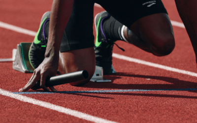
Sprinter set in the starting blocks on a red athletics track