opusbiological, Author at opusbiological.com - Page 3 of 9
Western Acupuncture and Dry Needling in the management of Musculoskeletal Conditions

Western Acupuncture and Dry Needling in the management of Musculoskeletal Conditions

Musculoskeletal (MSK) conditions can significantly impact an individuals’ quality of life i.e. through chronic pain, injury, muscle spasm or tightness. These MSK conditions in turn can then have a significant impact on mental health through anxiety and depression. Depression is four times more prevalent in people who suffer from persistent pain compared to those without (Gov.uk)

A recent analysis of Global Burden of Disease (GBD) 2019 data showed that approximately 1.71 billion people globally live with musculoskeletal conditions, including low back pain, neck pain, fractures, other injuries, osteoarthritis, amputation and rheumatoid arthritis (WHO). This will undoubtedly have  a huge impact on mobility, well-being, work attendance and therefore retirement age as well as people’s ability to participate in society.

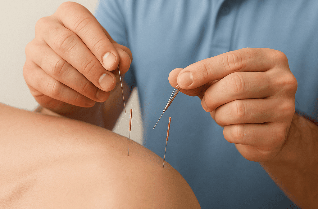
Western acupuncture is based on modern anatomical and physiological principles rather than those used in traditional Chinese Acupuncture. Single-use, sterile needles are used to stimulate specific points on the body to help alleviate pain and enhance function. Dry needling uses a similar concept and the same needles, but targets myofascial trigger points in order to help relieve tension. Chinese acupuncture which was first documented in China over 3000 years ago is thought to balance the flow of qi (energy) throughout your body, through the release of endorphins (Kirchhof-Glazier D 2014). Both Dry needling and acupuncture help to produce an inflammatory reaction, stimulating your body’s natural ability to heal (BMJ 2009).

Recent studies have provided substantial evidence demonstrating the effectiveness of Western acupuncture and dry needling in managing MSK conditions. A systematic review by Tough et al. (2021) indicates that both therapies significantly reduce pain and improve function in various musculoskeletal disorders. Their findings revealed notable reductions in pain intensity and enhancements in mobility following treatment (Tough et al., 2021).

In a randomized controlled trial, Cummings et al. (2022) examined the effects of dry needling on chronic shoulder pain. The results indicated that participants who received dry needling experienced a significant decrease in pain levels and improved shoulder function compared to a control group not receiving treatment.

The mechanisms underlying the effectiveness of both western acupuncture and dry needling involve the stimulation of the nervous system, which promotes the release of endorphins and other neurotransmitters helping to mitigate pain. This neurophysiological response not only alleviates immediate discomfort but may also facilitate long-term healing through enhanced blood flow to affected areas (Dyer et al., 2023).

Additionally, acupuncture and dry needling can induce muscle relaxation and restore proper movement patterns. This is particularly beneficial for conditions such as myofascial pain syndrome, characterized by tight muscles and trigger points. A study by Lee et al. (2020) demonstrated that patients undergoing dry needling exhibited significantly reduced muscle stiffness and improved range of motion, leading to an enhanced overall quality of life (Lee et al., 2020).

The safety of Western acupuncture and dry needling is another significant advantage. When administered by trained professionals, the risks associated with these treatments are minimal, making them suitable for a wide range of patients. A review by Johnson et al. (2022) reported that adverse effects are generally mild, such as temporary soreness or bruising at the needle sites, with serious complications being rare. It is also reported that  when combined with physiotherapy, chiropractic care, or other modalities, overall treatment outcomes for MSK conditions can be enhanced (Johnson et al., 2022).

In conclusion, Western acupuncture and dry needling present promising benefits for the management of musculoskeletal conditions. With a growing body of evidence supporting their effectiveness and safety, these therapies can be valuable additions to treatment options.

Reference List


  • Cummings, T. M., et al. (2022). “Efficacy of dry needling in the management of chronic shoulder pain: A randomized controlled trial.” Journal of Musculoskeletal Pain, 30(2), 89-96.
  • Dyer, D. L., et al. (2023). “The neurophysiological mechanisms of acupuncture: A review of the evidence.” Acupuncture in Medicine, 41(1), 15-22.
  • Johnson, C. D., et al. (2022). “Safety and efficacy of acupuncture and dry needling in the treatment of musculoskeletal disorders: A systematic review.” Physical Therapy Reviews, 27(4), 230-240.
  • Lee, J. H., et al. (2020). “Effects of dry needling on muscle stiffness and range of motion: A systematic review.” Journal of Bodywork and Movement Therapies, 24(4), 328-335.
  • Tough, E. A., et al. (2021). “The efficacy of acupuncture for musculoskeletal pain: A systematic review and meta-analysis.” European Journal of Pain, 25(7), 1345-1359.

Importance of calf capacity

Importance of calf capacity

The Importance of Good Calf Muscle Capacity in Preventing and Rehabilitating Lower Limb Injuries

Calf muscles, primarily consisting of the gastrocnemius and soleus muscles, play a vital role in lower limb function and overall biomechanics. These muscles are essential for movements such as walking, running, and jumping, contributing to ankle stability, propulsion, and shock absorption. Strong and flexible calf muscles not only enhance athletic performance but also prevent lower limb injuries and support rehabilitation after injury. This article outlines the importance of calf muscle capacity, provides data on its impact on joint loading, and explains how to assess calf capacity in clinical settings.

The Role of Calf Muscles in Lower Limb Function

The calf muscles work as a dynamic powerhouse during movement. The gastrocnemius and soleus muscles, in conjunction with the Achilles tendon, drive plantarflexion, which is the action of pushing the foot off the ground. This movement is crucial for walking, running, and jumping, as it propels the body forward. Additionally, the calf muscles play a role in absorbing the impact forces generated when the foot strikes the ground, thus reducing stress on other structures like the knee, hip, and lower back.

By acting as shock absorbers and stabilizers, the calf muscles influence the load distribution across the entire lower limb. Proper calf function ensures that forces are dispersed evenly, preventing overloading of other structures and contributing to balanced biomechanics. When calf muscles are weak or fatigued, they fail to perform this role efficiently, leading to improper movement patterns and potentially contributing to injuries of the lower limb, such as ankle sprains, Achilles tendinopathy, anterior knee pain, and plantar fasciitis. Weak calf musculature may also exacerbate symptoms such as osteoarthritis in the foot, ankle and knee.  A study by Alshami and Alhassany (2020) 1, found that calf muscle strength was significantly reduced in the patients with knee OA compared with that in the healthy control participants. Therefore it could be a good idea to ensure that within an OA knee patient’s exercise programme, there is targeted calf strengthening.

Impact of Strong Calf Muscles on Knee Joint Loading

Strong calf muscles not only support the ankle but also significantly reduce the load on the knee joint during various activities. Research has shown that adequate calf strength can reduce knee joint loading by absorbing the ground reaction forces that occur during walking or running. The soleus muscle, in particular, is known to play a key role in reducing the stress on the knee by generating posterior shear forces at the tibia, which counteract the forward forces that tend to overwork the knee.

A 2019 study by A. Bohm, 2,  found that the soleus muscle can contribute up to 50% of the force needed to decelerate the body during walking and running, thereby offloading the knee joint considerably. Research indicates that strong calf muscles can reduce the load on the knee by up to 20-25% during activities like walking and stair climbing . This effect is critical in both preventing injuries such as patellofemoral pain syndrome and in managing conditions like osteoarthritis, where reduced joint loading can slow the progression of cartilage wear.

In individuals with weakened calf muscles, this offloading effect is diminished, forcing the knee and other structures to take on additional load, which increases the risk of injury. Athletes, in particular, benefit from strong calf muscles, as these muscles allow them to handle high-impact activities like running and jumping without overstressing the knee joint.

Testing Calf Capacity in Clinical Settings

Given the importance of calf muscle capacity in preventing and rehabilitating lower limb injuries, clinicians need reliable methods to assess both the strength and endurance of the calf muscles. Below are some of the most effective ways to test calf capacity in a clinical setting:

  • Calf-Raise Test (Repetitive Heel-Raise Test): The single-leg calf-raise test is a simple and widely used method to assess calf muscle endurance. In this test, the patient stands on one leg and performs heel raises, lifting the heel off the ground as high as possible with the number of repetitions completed before fatigue, recorded. This test evaluates both the gastrocnemius and soleus muscles, providing a measure of endurance, which is critical for injury prevention. Hebert et. Al (2017) 2, carried out a great study which generated a guide to target rep ranges for different patient groups based on age and sex.
  • Maximal Voluntary Isometric Contraction (MVIC): This test measures the maximum force a person can generate during an isometric contraction of the calf muscles. Using a dynamometer, clinicians can measure the force output when the patient pushes against resistance in a fixed position. The MVIC is a reliable method to assess the maximal strength of the calf muscles, especially useful in identifying deficits that may contribute to injury risk or delay rehabilitation.
  • Single-Leg Hop Test: This test is used to evaluate functional strength and power in the calf muscles. The patient is asked to perform a series of single-leg hops for distance or height. A significant discrepancy between the two legs indicates potential calf muscle weakness or imbalance. This test is particularly useful for athletes who need to perform explosive movements during sport, as it mimics the high-force demands placed on the calf muscles.
  • Functional Movement Assessments: Assessing how the calf muscles function during movements like squats, walking, or running is also valuable in clinical settings. Analysing gait or performing functional movement screens allows clinicians to observe whether the calf muscles are functioning optimally during dynamic activities. This type of assessment is particularly relevant for athletes and individuals recovering from injury, as it highlights any biomechanical deficiencies related to calf muscle performance.

Rehabilitation of Lower Limb Injuries: Calf Strengthening

Injury rehabilitation programs often include exercises aimed at restoring calf muscle strength, endurance, and flexibility. Isometric calf exercises can be used early in rehabilitation to improve muscle activation without placing too much strain on the injured tissue. Over time, patients progress to more dynamic exercises such as calf raises, hopping, and plyometric drills, depending on the severity of their injury and their physical goals. Green et. Al (2022) 3, break up the involvement of calf specific exercises into 4 nice stages which a patient or player should go through in order to go from day 1 to return to play. These stages are: Early loading and foundation calf and lower limb function, Loaded strengthening, loaded power, plyometrics and ballistics and finally, Locomotion.

Conclusion

Good calf muscle capacity is vital for preventing lower limb injuries and supporting effective rehabilitation. Strong and flexible calf muscles reduce the load on the knee joint by as much as 20-25%, play an essential role in stabilizing the ankle, and help prevent common injuries such as Achilles tendinopathy, plantar fasciitis, and knee injuries. Clinicians can use a variety of tests, such as the calf-raise test and MVIC, to assess calf muscle strength and endurance. By incorporating calf training and flexibility exercises into rehabilitation and prevention programs, individuals can improve their lower limb function, reduce the risk of injury, and ensure a quicker recovery following injury.

Reference List


  • Alshami, A.M. and Alhassany, H.A. (2020). Girth, strength, and flexibility of the calf muscle in patients with knee osteoarthritis: A case–control study. Journal of Taibah University Medical Sciences, 15(3), pp.197–202. doi:https://doi.org/10.1016/j.jtumed.2020.04.002.
  • Bohm S, Mersmann F, Santuz A, Arampatzis A.. 2019. The force–length–velocity potential of the human soleus muscle is related to the energetic cost of running. Proc. R. Soc. B Biol. Sci. 286, 20192560 ( 10.1098/rspb.2019.2560)
  • Hébert-Losier, K., Wessman, C., Alricsson, M. and Svantesson, U. (2017). Updated reliability and normative values for the standing heel-rise test in healthy adults. Physiotherapy, 103(4), pp.446–452. doi:https://doi.org/10.1016/j.physio.2017.03.002.
  • Green, B., McClelland, J.A., Semciw, A.I. et al. The Assessment, Management and Prevention of Calf Muscle Strain Injuries: A Qualitative Study of the Practices and Perspectives of 20 Expert Sports Clinicians. Sports Med – Open 8, 10 (2022). https://doi.org/10.1186/s40798-021-00364-0

Injuries in paddle

Injuries in paddle

Paddle tennis has increased in popularity over the last few years and due to the fast-nature of the sport, it is unsurprising that musculoskeletal injuries are very common. Paddle takes place on a small field and surrounded by glass which increases the speed of the game. Paddle requires ‘motor anticipation, explosive force, reactive speed (acceleration and deceleration), resistance, flexibility and agility’(Virgolino, 2022). These components of fitness are key for health, wellness and longevity however there is an increased risk of injury if one does not apply appropriate preventative techniques (Virgolino, 2022).


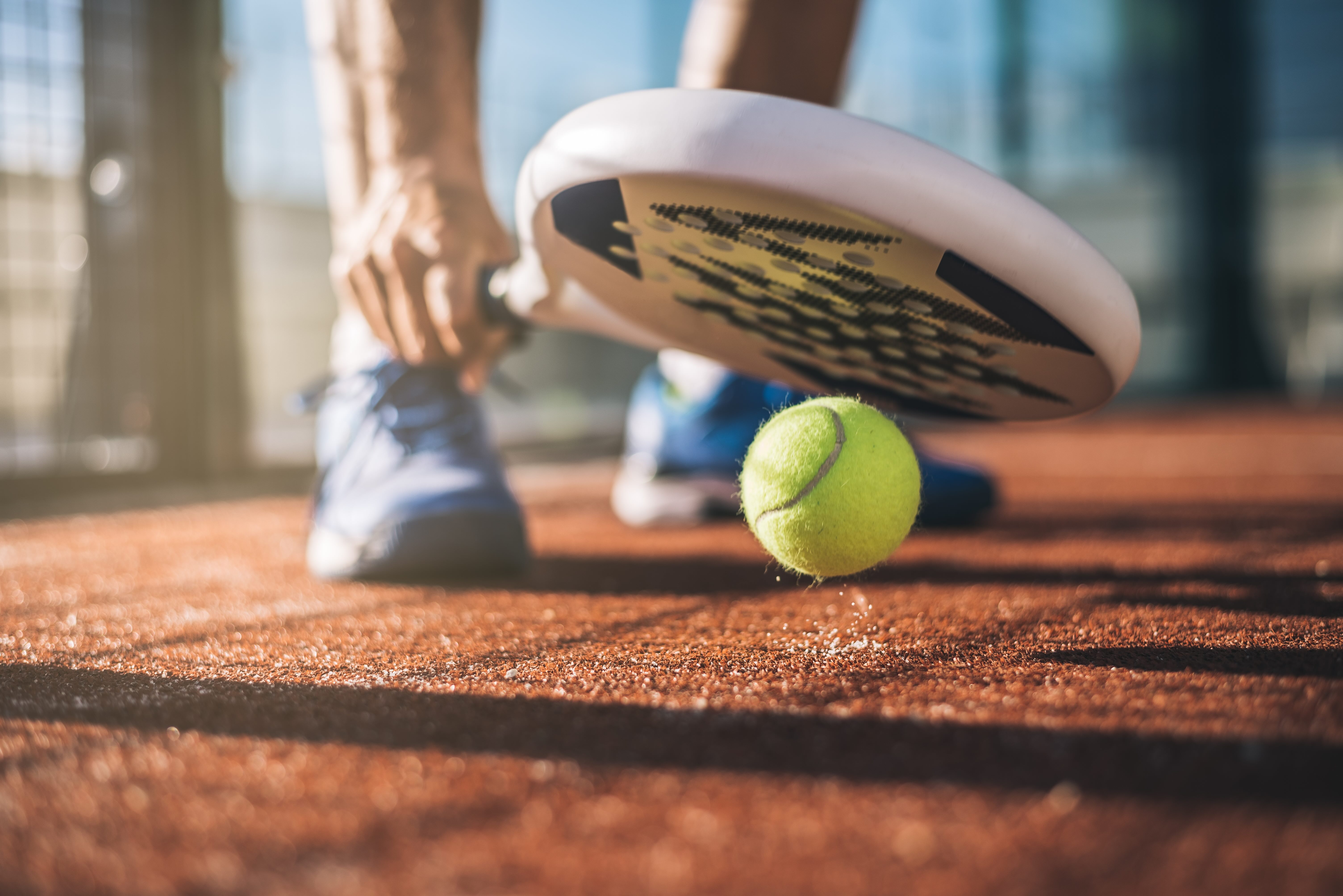
Sportsman playing padel game

The most common MSK injuries in senior players include knee sprains, lateral epicondylitis, shoulder pain (20.6%) and lower back pain (27.5%) (Castillo-Lozano, 2017). However, lower back pain is the most prevalent among junior players (Virgolino, 2022). These findings enable practitioners to create preventive strategies and programmes which focus on scapular stability, trunk stability and lower limb strength (Castillo-Lozano, 2017).  Despite the risk of injury, paddle provides many benefits, especially to elderly players. There is a variety of research demonstrating the physical advantages (muscular strength, endurance and reaction time) of paddle, however literature has shown paddle to also provide psychological benefits such as reducing anxiety and stress (Eirale, 2023).

Elbow tendinopathy mostly impacts novice players. This is most likely due to players hitting backhand strokes in wrist flexion, the sudden increase of eccentric loading of forearm extensors and the vibrations impacting the elbow when the ball reaches the racket. Lateral epicondylitis is extremely common due to the repetitive movements and duration of the game (2-4hours). However there have been recent improvements in racket technology which reduce the risk of epicondylitis.  It is also unsurprising that patella tendinopathy often occurs due to repetitive jumping and sudden change in direction. Interestingly, males are 2-4 times more likely to develop patella tendinopathy, whereas females are more likely to experience chondromalacia patellae (Eirale, 2023).

Disc-related lower back pain and facet joint syndrome are predominantly the main cause of lower back pain (LBP) in paddle. Sward et. al found 50% of their participants suffered with LBP and 1/5 experienced severe symptoms. This is likely due to paddle incorporating all ranges of motion in the lumbar spine combined with explosive movements; therefore, it is important to consider trunk stability and lumbar mobility to prevent injury (Eirale, 2023).

Lastly, repetitive movements such as ‘smash’ and ‘bandejas’ cause muscle imbalances in the shoulder, increasing the risk of rotator cuff injuries and scapular instability. Also due to repeated overhead movements, there is a possibility of impingement in the glenoid humeral joint leading to a SLAP lesion. Consequently, rotator cuff strengthening and scapular stability are key to prevent shoulder injuries (Eirale, 2023).

Reference list

Castillo-Lozano, R. (2017). Epidemiology and prevention strategies for the musculoskeletal injuries in the paddle-tennis senior players. Science & Sports, 32(3), pp.e101–e106. doi:https://doi.org/10.1016/j.scispo.2016.09.009.

Eirale, C. (2023). Aspetar Sports Medicine Journal – INJURIES IN PADEL. [online] Aspetar.com. Available at: https://journal.aspetar.com/en/archive/volume-12-targeted-topic-sports-medicine-in-padel/injuries-in-padel [Accessed 22 Aug. 2024].

Virgolino, M. (2022). Running in Paddle. Springer eBooks, pp.179–183. doi:https://doi.org/10.1007/978-3-662-65064-6_19.


Stay Active, Stay Healthy: The Importance of Physiotherapy for Active Individuals

Stay Active, Stay Healthy: The Importance of Physiotherapy for Active Individuals

Maintaining an active lifestyle is crucial for overall health and well-being in today’s fast-paced world. Whether you’re a seasoned athlete or simply someone who enjoys staying active through sports and exercise, taking care of your body is paramount. However, as you age, changing your approach to fitness is essential to stay active. Proactive preventive approaches such as physiotherapy can prevent injuries, enhance performance, and enable you to continue enjoying the activities you love for years to come.

Who is a physiotherapist? How can a physiotherapist help me?

Physiotherapists, also known as physical therapists, are highly trained healthcare professionals who specialize in assessing, diagnosing, and treating a wide range of musculoskeletal and movement-related conditions. Their primary focus is on optimizing movement, promoting physical well-being, preventing injuries, and rehabilitating individuals who have experienced physical impairments. Here’s an overview of what physiotherapists do and how they can help you stay active:
  • Assessment: Physiotherapists begin by comprehensively assessing the patient’s condition. This involves evaluating their physical function, range of motion, strength, posture, and pain or discomfort. The goal is to identify the underlying issues that may be causing the patient’s problems.
  • Diagnosis: Based on the assessment, physiotherapists diagnose the underlying causes of the patient’s symptoms. They determine the root of the issue, whether it’s a specific injury, a musculoskeletal problem, a neurological condition, or a combination of factors.
  • Treatment Planning: Physiotherapists create personalized treatment plans tailored to the patient’s needs, goals, and condition. These plans often include a combination of various therapeutic techniques and exercises designed to address the identified issues.
  • Exercise Prescription: Physiotherapists prescribe specific exercises and therapeutic activities that target the patient’s condition. These exercises strengthen weak muscles, improve flexibility, enhance balance, and address movement abnormalities.
  • Patient Education: Educating the patient about their condition, proper body mechanics, injury prevention, and self-management techniques is a significant part of a physiotherapist’s role. Patients are empowered to participate in their recovery and long-term health actively.
  • Rehabilitation: Physiotherapists play a crucial role in the rehabilitation process after surgeries, accidents, or injuries. They help patients regain strength, flexibility, and function, guiding them through a structured recovery program.
  • Monitoring Progress: Throughout the treatment process, physiotherapists continually assess the patient’s progress and adjust the treatment plan as needed to ensure optimal results.

Top 5 reasons why physiotherapy is essential for active individuals

Active lifestyles put increased stress on our bodies, making them more susceptible to overuse injuries, muscle imbalances, and joint problems. Physiotherapists at Opus Biological specialize in identifying and addressing these issues before they escalate, thus reducing the risk of injuries. They design personalized exercise plans to strengthen weak areas, improve flexibility, and optimize biomechanics, improving performance and efficiency in sports and activities. Moreover, as individuals age, physiotherapy ensures that they can adapt their exercise routines to maintain mobility, balance, and function, allowing them to enjoy an active lifestyle for life. Here are the top 5 reasons to choose physiotherapy to maintain an active lifestyle:

  • Injury Prevention: Active individuals often engage in high-impact activities that put a strain on muscles, joints, and ligaments. Physiotherapy helps identify areas of weakness, imbalance, or poor biomechanics. Through targeted exercises and corrective techniques, physiotherapists can prevent injuries by strengthening these vulnerable areas, reducing the risk of overuse injuries, strains, and sprains.
  • Improved Performance: Physiotherapy isn’t just about preventing injuries; it’s also about optimizing performance. Physiotherapists can analyse an individual’s movement patterns, identify inefficiencies, and provide tailored exercises to improve form, strength, and flexibility. This optimization leads to better athletic performance and increased endurance.
  • Faster Recovery: If an active individual does experience an injury, physiotherapy is essential for a swift and effective recovery. Physiotherapists employ manual therapy, targeted exercises, and modalities like ultrasound or electrical stimulation to speed up the healing process, restoring function and reducing the risk of complications.
  • Personalized Guidance: Physiotherapy at Opus Biological offers individualized care that aligns with your needs, goals, and physical condition. This personalized approach ensures that active individuals receive tailored advice, exercises, and strategies that address their unique challenges, helping them achieve their fitness objectives while minimizing the risk of setbacks.
  • Staying Active for Life: Physiotherapy helps adapt exercise routines to address age-related changes, such as reduced joint flexibility or muscle strength. This ensures active individuals can continue enjoying their preferred activities as they age, promoting overall health and well-being.

Managing Injuries Better with Physiotherapy

Preventing injuries is always better than treating them, and that’s where physiotherapy shines. A physiotherapist is a healthcare professional trained to assess and address musculoskeletal issues, identify potential problems, and design personalized prevention plans. By identifying weak areas, poor movement patterns, or imbalances, a physiotherapist can create exercises and strategies to address these issues, reducing the risk of injury.

For active individuals, this proactive approach is a game-changer. It allows you to identify and correct small issues before they become significant problems. It’s like giving your body a regular check-up, ensuring every joint, muscle, and ligament functions optimally. By investing time in physiotherapy, you’re investing in your body’s longevity.

Individualized Care Matters

One of the significant advantages of physiotherapy at Opus Biological is its personalized approach. Every person is unique, and their needs, goals, and physical conditions are distinct. Our physiotherapists work closely with you to understand your specific conditions and design a customized plan that addresses your concerns.
Whether recovering from a sports injury, looking to improve your athletic performance, or seeking guidance on maintaining an active lifestyle as you age, our physiotherapists can provide the tailored care you require. The individualized attention you receive ensures that you’re on the right path, with exercises and strategies that align with your goals, ultimately helping you achieve desired results.

Final Thoughts

Staying active is a beautiful way to maintain your health and enjoy life to the fullest. However, taking a proactive approach is essential to ensure that you can continue being active without unnecessary setbacks. Physiotherapy offers a comprehensive solution, focusing on prevention, performance enhancement, and longevity in your active lifestyle.
By investing in regular physiotherapy sessions at Opus Biological, you’re investing in your body’s well-being. You’re taking steps to prevent injuries, optimize your performance, and adapt your approach to physical activity as you age. With the guidance of a skilled physiotherapist, you can ensure that you’re staying active and healthy for years to come, enabling you to enjoy the activities you love and lead a fulfilling, active life. So, make physiotherapy an integral part of your active lifestyle – your body will thank you for it.
Importance of calf capacity

The Effectiveness of Physiotherapy on Subacromial Impingement Syndrome

Subacromial impingement syndrome (SAIS) is an umbrella term for a variety of inflammatory pathologies within the subacromial space. These include rotator cuff syndrome, bursitis, tendinopathy and calcific tendinitis. Conservative treatment is used to decrease inflammation, restore range of motion in the shoulder and strengthen the rotator cuff (Gebremariam et. al, 2013). Physiotherapy has shown to be an effective method for treating SAIS and should be considered as the first line of care (Dicken et. al, 2005). However, it is important to understand which therapeutic modalities should be prioritised and considered when treating SAIS.

There is a wealth of research suggesting to focus on scapular stabilization and rotator cuff strengthening when rehabilitating patients with SAIS. For example, Baskurt et. all investigated the effectiveness of scapular stabilization on pain, shoulder range of motion, joint position sense (JPS), scapular dyskinesis and quality of life in patients with SAIS. They discovered the combination of scapular stabilization, shoulder strengthening and stretching is the most effective method in improving JPS and decreasing scapular dyskinesis (Baskurt et. al, 2011).  Michener et. al compared various treatments for SAIS (exercise, joint mobilisations, laser, ultrasound and acupuncture) in a systematic review. The evidence suggested laser therapy is only beneficial when used in isolation and there is no benefit in using ultrasound or acupuncture. However, the most effective treatment for SAIS is the combination of rotator cuff strengthening and scapular stabilisation alongside joint mobilisations (Michener et. al, 2004). Additionally, Celik concluded that patients with SAIS exhibited a lower visual analog scale score when exercises were performed within pain free range of motion (90 degrees and below). Overall research illustrates rotator cuff strengthening, scapular stabilization and joint mobilization are essential when treating SAIS. Furthermore, during the early stages of rehabilitation, prescribing exercises below 90-degree shoulder flexion can aid in pain management (Celik, 2009).

When creating a rehabilitation programme, practitioners should consider a patient-centered approach. It is important to recognize patients’ pain tolerance and threshold when prescribing exercises for SAIS. Vallés- Carrascosa et. al compared painful eccentric exercises (PEE) to pain free eccentric exercises when treating SAIS. Remarkably, PPE was found to provide no additional benefits. Therefore, practitioners should consider prescribing pain free exercises to promote compliance with rehabilitation programmes, especially for patients with a lower pain tolerance (Vallés- Carrascosa et. al,2018).


Reference List


  • Başkurt, Z., Başkurt, F., Gelecek, N. and Özkan, M.H. (2011). The effectiveness of scapular stabilization exercise in the patients with subacromial impingement syndrome. Journal of Back and Musculoskeletal Rehabilitation, 24(3), pp.173–179. doi:https://doi.org/10.3233/bmr-2011-0291.
  • Celik, D. (2009). Comparison of the effects of two different exercise programs on pain in subacromial impingement syndrome. Acta Orthopaedica et Traumatologica Turcica, 43(6), pp.504–509. doi:https://doi.org/10.3944/aott.2009.504.
  • Dickens, V.A., Williams, J.L. and Bhamra, M.S. (2005). Role of physiotherapy in the treatment of subacromial impingement syndrome: a prospective study. Physiotherapy, 91(3), pp.159–164. doi:https://doi.org/10.1016/j.physio.2004.10.008.
  • Gebremariam, L., Hay, E.M., van der Sande, R., Rinkel, W.D., Koes, B.W. and Huisstede, B.M.A. (2013). Subacromial impingement syndrome—effectiveness of physiotherapy and manual therapy. British Journal of Sports Medicine, 48(16), pp.1202–1208. doi:https://doi.org/10.1136/bjsports-2012-091802.
  • Michener, L.A., Walsworth, M.K. and Burnet, E.N. (2004). Effectiveness of rehabilitation for patients with Subacromial impingement syndrome: a systematic review. Journal of Hand Therapy, 17(2), pp.152–164. doi:https://doi.org/10.1197/j.jht.2004.02.004.
  • Vallés-Carrascosa, E., Gallego-Izquierdo, T., Jiménez-Rejano, J.J., Plaza-Manzano, G., Pecos-Martín, D., Hita-Contreras, F. and Achalandabaso Ochoa, A. (2018). Pain, motion and function comparison of two exercise protocols for the rotator cuff and scapular stabilizers in patients with subacromial syndrome. Journal of Hand Therapy, 31(2), pp.227–237. doi:https://doi.org/10.1016/j.jht.2017.11.041.