opusbiological, Author at opusbiological.com
What the Women’s Euros Teach Us: Lionesses Win, But There’s More to Learn About Injury in the Women’s Game

What the Women’s Euros Teach Us: Lionesses Win, But There’s More to Learn About Injury in the Women’s Game

On Sunday 27 July 2025, England’s Lionesses retained their European crown in thrilling fashion, defeating Spain 3–1 on penalties after a tense 1–1 draw at St. Jakob‑Park in Basel. It was a performance defined by resilience, tactical adaptability, and mental steel.

But while England’s back-to-back UEFA Women’s Euro victories rightly dominate the headlines, an equally important story unfolds. The growing need to address sex-specific injury risks in female footballers, especially as the women’s game continues its rapid ascent.

England’s Comeback Queens

Despite trailing 0–1 at half-time, England mounted a spirited second-half comeback, sparked by substitute Chloe Kelly, who delivered the cross for Alessia Russo’s 57th‑minute equaliser. With no goals in extra time, the match moved to penalties, where Kelly once again became a national hero, burying the final spot-kick and sealing England’s first major title won abroad.

  • “They know how to win, they had proven it before, and that was all they needed to turn to in the toughest moments.” – BBC Sport

https://www.bbc.co.uk/sport/football

Statistically, the Lionesses defied the odds throughout Euro 2025: https://www.uefa.com/womenseuro/statistics/

  • Came from behind in all three knockout matches
  • Had 10 goal involvements from substitutes, a tournament record
  • Became the first team to win the final after trailing at half-time

But resilience often comes at a price. Particularly when it comes to the physical toll on players’ bodies.

The Injury Disparity Between Male and Female Footballers

One of the starkest issues in elite women’s football is the prevalence of non‑contact injuries, especially anterior cruciate ligament (ACL) tears. Women are estimated to be 6–8 times more likely than men to suffer ACL injuries in football due to anatomical, hormonal and biomechanical factors.

A 2021 study published in the British Journal of Sports Medicine observed that: https://bjsm.bmj.com/content/55/3/135

  • “Female athletes demonstrate altered landing mechanics, greater valgus knee angles, and hormonal fluctuations that increase ligament laxity, particularly during ovulation.”

And this isn’t just theory. Lucy Bronze reportedly played the entire tournament with a stress fracture, while multiple squads have quietly battled ongoing muscular and ligament injuries that disproportionately affect women at this elite level.

Why Women Need Tailored Sports Medicine

Despite progress, many training regimes remain based on male physiology. They often overlook the complexities of the female athlete’s endocrine system, injury profile and recovery curve.

Research, including the 2024 UEFA Women’s Health Report https://www.uefa.com/insideuefa/news/0278-15ea58b9fdd7-c84169b43d5e-1000–women-s-football-and-health-report-2024/, emphasises the urgent need to adapt everything from pre‑season screening and load management to menstrual‑cycle tracking and neuromuscular conditioning for ACL prevention. Yet only a minority of professional clubs have fully integrated female-specific health monitoring into their high-performance frameworks.

We believe this must change. At Opus we are proud to lead that transformation.

Regenerative Therapies and Prevention at Opus

To meet the needs of today’s elite female athletes, Opus offers a holistic blend of sports medicine and rehabilitation:

Service  Description
Sports Medicine Expert prevention, diagnosis and rehab tailored to musculoskeletal injuries and performance optimisation.
Regenerative Medicine Including allogeneic umbilical cord-derived mesenchymal stem cell therapy and platelet-rich plasma (PRP) injections, integrated with bespoke rehab programmes.
Reformer Pilates For core stability, neuromuscular control and injury prevention.
ACL Prevention Programmes Dedicated protocols to reduce ACL risk via targeted neuromuscular training.
Menstrual Cycle-Informed Training Protocols Tailored load management and timing based on hormonal cycles.

We treat athletes not just based on the injury, but on their unique physiological and hormonal context. This creates personalised pathways to longevity and peak performance.

Final Whistle: Lessons Beyond the Pitch

England’s Euro 2025 victory is a testament to elite preparation, adaptability and belief. But it also reminds us that women’s football is entering a new phase of professionalism. One that demands evolving our understanding of injury risk, prevention and care for female athletes.

As we celebrate the Lionesses’ glory, let’s also commit to building systems that keep women stronger, longer – on the pitch and beyond.

Want to Futureproof Your Athletic Health?

Whether you’re a professional athlete or striving for optimal performance, Opus offers the city’s most advanced destination for sports injury prevention and recovery. 

Whether you’re a professional athlete or striving for optimal performance, Opus offers the city’s most advanced destination for sports injury prevention and recovery. 

Book a consultation with Dr David Porter or our multi-disciplinary team today.

📍 Located in the heart of London
📞 Call us on [020 8609 7843]

Dr David Porter’s Perspective on Tennis Injuries at Wimbledon – and How to Stay on Court

Dr David Porter’s Perspective on Tennis Injuries at Wimbledon – and How to Stay on Court

I’m Dr David Porter, Sports & Exercise Medicine physician at Opus, and former first team doctor to Chelsea football club (https://www.chelseafc.com/en). Each June the lawns of the All England Club become the focal point of the tennis world at Wimbledon (https://www.wimbledon.com/index.html)​​, but for clinicians like me the tournament also highlights the epidemiology of overuse and acute injuries that recreational players will mirror on the municipal courts a week later. Below I outline the most common conditions I diagnose in tennis enthusiasts, the underlying biomechanics, and the integrated prevention‑and‑treatment strategies we employ at Opus.

Quick read: If you’re in pain now, book an assessment with our physiotherapy team via the Opus website or call  +44 20 8609 7843 – prompt diagnosis limits time off court.

The Physical Demands of Modern Tennis

The modern baseline‑dominated game generates peak rotational velocities exceeding 4,000°/s during the forehand¹, placing tremendous eccentric load on the wrist extensor‑supinator complex. Matches can last over three hours, requiring repeated accelerations and decelerations that stress every major joint.

Key reference data:

  • Serve speeds: up to 140 mph according to the International Tennis Federation (ITF).
  • Change‑of‑direction events: 600–1,000 per five‑set match (source: British Journal of Sports Medicine).
  • Annual incidence of injury: 2.3–3 injuries per 1,000 playing hours in amateurs per the Lawn Tennis Association(LTA).

Common Tennis Injuries and Conditions

Lateral epicondylitis (“Tennis Elbow”)

  • Pathophysiology: Degenerative tendinopathy of the extensor carpi radialis brevis.
  • Risk factors: Excess grip size, late‐contact backhands, and sudden string‑tension changes – see the NHS tendonitis guidance.
  • Presentation: Lateral elbow pain on resisted wrist extension, weak grip.
  • Treatment pathway:
    1. Relative rest & Load Modification (POLICE principle) – NICE CKS.
    2. Eccentric‑concentric exercise supervised by our physios.
    3. Shock‑wave therapy – systematic review in BJSM.
    4. Platelet‑Rich Plasma (PRP) or Mesenchymal Stem Cell (MSC) injections where refractory, following protocols described in Regenerative Medicine.

Shoulder Impingement & Rotator‑Cuff Tendinopathy

High‑velocity serves predispose to internal impingement, supraspinatus overload and posterior capsular tightness.

  • MRI evidence of humeral retroversion in players is detailed in The American Journal of Sports Medicine.
  • My algorithm emphasises scapular control drills and isoinertial rotator‑cuff loading, informed by Quatman et al. 2024.
  • For labral tears or partial cuff tears I liaise with our shoulder surgeon colleagues.

Lumbar Stress Injuries

Lumbar extension and axial rotation during the serve create shear forces up to 1.7 body‑weights².

Patellar & Quadriceps Tendinopathies

Jumping and deceleration provoke patellar tendon strain. Risk rises on grass due to low friction requiring deeper knee flexion.

  • Prevention: Nordic hamstring and single‑leg decline squat programmes (see Physio‑Pedia).
  • Therapies: Isolated heavy–slow‑resistance (HSR) loading, PRP, and in recalcitrant cases ultrasound‑guided MSC injection following the BrIT Therapy consensus.

Ankle Inversion Sprains

Grass surfaces increase slip‑risk. The majority are ATFL grade I–II.

  • Immediate care: POLICE with early weight‑bearing — as per NICE NG59.
  • Rehabilitation: Sensorimotor control work on the Pilates reformer’s unstable carriage, and late‑stage return‑to‑sport drills based on FIFA 11+.
  • Re‑injury reduction: Semi‑rigid ankle braces; meta‑analysis in Sports Medicine‑Open.

Hand Blisters & Calluses

Often dismissed, blisters can derail championships (cf. Agassi 1996). We use Dermabond adhesive and grip‑tapings from the ITF Medical Commission.

Heat‑Related Illness & Hydration‑Electrolyte Imbalance

Even in London, Centre Court’s micro‑climate can reach 30 °C. Follow the NCAA Heat Policy and the “3 Cs” I teach: Colour of urine, Change in body‑mass <2%, Consistency of intake (sipping not gulping).

Diagnostic Approach at Opus

  1. History & Biomechanical Screen 
  2. Point‑of‑care ultrasound for tendon and ligament evaluation
  3. MRI

Evidence‑Based Treatment Options

Modality

Evidence Level

Typical Indications

Notes

Physiotherapy (mechanotherapy)

1A (Cochrane 2025)

Most overuse injuries

Individualised loading programmes

Shockwave Therapy

1B

Chronic tendinopathy

Radial or focused waves

Corticosteroid Injection

1B but short‑term

Acute bursitis, impingement

Counsel about transient gain

PRP Injection

2B

Refractory epicondylitis

Double‑spin leukocyte‑poor

MSC Therapy

2C emerging

Partial cuff tears, tendinopathy >6 mo

Discuss ongoing trials (ClinicalTrials.gov)

Reformer Pilates

2B

Lumbo‑pelvic stability

Opus‑designed tennis‑specific protocol

Custom Orthoses

1C

Pes cavus with lateral ankle sprains

Casted in neutral subtalar

Levels according to the Oxford CEBM.

Prevention Strategies That Work

Dynamic Warm‑Up

Adopt “RAMP” (Raise–Activate–Mobilise–Potentiate) protocols (see UK Sport). Five minutes of skipping, leg swings, inchworms and resisted band external rotation reduces soft‑tissue injury by up to 30%.

Strength & Conditioning

I prescribe:

  • Eccentric wrist extensor loading — protocol from Stasinopoulos & Manias.
  • Rotator‑cuff HSR in 15° scapular plane abduction — guidelines at Physiopedia.
  • Eccentric–isometric patellar control (Dye’s quadrant approach).

Equipment Tuning

  • Grip Size: Spencer et al. showed 15% grip‑size error increases extensor torque by 25% (Journal of Hand Surgery).
  • String Tension & Gauge: Lower tensions (<55 lbs) reduce peak shoulder internal rotation moment by 6% (ITF Technical Centre).
  • Footwear: Anti‑torsion shank and grass‑court outsole patterns minimise inversion loads; research in Foot wear Science shows that a pronounced lateral‑edge flare can reduce peak eversion torque by 12 %. 

Footwork & Neuromuscular Agility

Efficient split‑steps and first‑step explosiveness are modifiable factors. Six weeks of ladder drills and reactive cone work improved time‑to‑stabilisation by 18 % in collegiate players in a randomised trial published in the Journal of Strength & Conditioning Research. We super‑set these with Pilates jump‑board intervals to integrate proximal hip stability, referencing principles from Polestar Education.

Tailoring Your Game to the Court Surface

Surface

Mechanical Demand

Injury Bias

Practical Tip

Grass (Wimbledon)

Low friction, low coefficient of restitution

Ankle inversion, knee extensor overload

Wider base during deceleration; use herring‑bone outsole pattern (ITF Surface Pace Ratings)

Clay

Higher friction, longer rallies

Adductor strains, lumbar rotation stress

Lunge conditioning and slide‑control drills; consult Rafa Nadal Academy methodology

Hard

High vertical GRF, predictable bounce

Patellofemoral pain, Achilles tendinopathy

Alternate insole densities; follow ACSM surface‑impact guidelines

Rotate footwear in line with the “Surface ⇄ Shoe ⇄ Load” model described by Silva et al. 2023.

Periodisation & Recovery – The Missing Set

“Most club players compete every week and train ad‑hoc – the opposite of what physiology dictates.”

Micro & Meso Cycles

  • Adopt a 3 : 1 load–deload ratio (three progressive weeks, one consolidation) as endorsed by the UK Strength & Conditioning Association.
  • Use session‑RPE × duration to monitor weekly Acute–Chronic Workload Ratio (ACWR); keep it 0.8‑1.3 (see Gabbett 2020).

Recovery Pillars

  1. Sleep: ≥7 h; use wearable metrics from WHOOP.
  2. Nutrition: 1.6‑1.8 g protein·kg⁻¹·day⁻¹; consult British Dietetic Association.
  3. Active recovery: 20 min low‑intensity cycling enhances venous return (study in European Journal of Applied Physiology.
  4. Cold‑Water Immersion: 11‑15 °C for 12 min lowers DOMS by 16 % — meta‑analysis in Sports Medicine.

Return‑to‑Play (RTP) Decision‑Making

A purely time‑based clearance is outdated. 

  1. Resolution of pain (VAS <1/10) and inflammation.
  2. Range of motion within 95 % of contralateral limb.
  3. Readiness testing — isokinetic strength deficit <10 % (via Biodex).
  4. Reactive agility equal to baseline on the Y‑Balance Test.
  5. Re‑integration — graduated match play under coach supervision.

We document objective criteria in the electronic medical record (EMR) and share it with the athlete’s coach and, when appropriate, with their LTA Accredited+ strength & conditioning professional.

Key Take‑Home Messages

  • Load management beats miracle cures. Every chronic tennis injury I treat began as a manageable ‘niggle’.
  • Technique tweaks ≈ tissue stress shifts.
  • Integrated care accelerates outcomes. 

Need help? 

Call +44 20 8609 7843 or

info@opusbiological.com

Same‑day appointments available.

Disclaimer

This blog post is for general educational purposes. It is not a substitute for personalised medical advice. If you have persistent pain or functional limitation, consult a qualified healthcare professional.

Role of Physiotherapy in Fibromyalgia

Role of Physiotherapy in Fibromyalgia

Fibromyalgia (FM) is a chronic condition which causes pain throughout the body. Other symptoms include fatigue, poor sleep quality, anxiety and depression (Antunes and Marques, 2022). The cause of chronic pain in patients with FM is associated with the hypersensitivity of the central nervous system resulting in hyperalgesia and potentially allodynia. The symptoms of fatigue and exhaustion can be explained by the hyporesponsiveness of the hypothalamus-pituitary- adrenal mechanisms in patients with FM (Mengshoel, 2013). Recently, more research is being conducted into the role of physiotherapy in FM and has shown to reduce pain, improve quality of life and function.

Exercise has been shown to improve psychological well-being and a reduction in pain, fatigue and other comorbidities (which is common in patients with FM). The combination of exercise and pain education have been proven to be the most effective intervention for FM (Mengshoel,2013) and also reduce medication consumption. Interestingly, addressing central sensitization and the neuroscience of pain in-person with patients has been shown to be the most successful form of pain education when improving compliance with exercise. However, there is insufficient data on what type of exercises is the most effective (Antunes and Marques, 2022).

Hydrotherapy and land exercise has been shown to be equally effective, although being immersed in water can have an analgesia effect. The heat and the increase of buoyancy causes the release of endorphins, increased capillarization and oxygen consumption. Moderate to high intensity resistance training has shown to reduce fear avoidance and improve neuromuscular ability which is especially important for menopausal and postmenopausal women. However, research demonstrates aerobic exercise to be more tolerated by patients (Antunes and Marques, 2022).

Physiotherapists regularly use massage and manual therapy for pain relief in patients with FM. Unfortunately, there are only a few studies addressing the effects of massage in FM (Antunes and Marques, 2022). Other modalities, such as TENS, have been proven to be effective in modifying pain and in turn an increased activity levels for patients with FM (Mengshoel, 2009).

There is conflicting evidence on the optimum treatment for patients with FM, however it is important to incorporate a multi-faceted approach that addresses the patients’ needs and goals.

Antunes, Mateus Dias, and Amélia Pasqual Marques. “The Role of Physiotherapy in Fibromyalgia: Current and Future Perspectives.” Frontiers in Physiology, vol. 13, no. 13, 16 Aug. 2022, p. 968292, www.ncbi.nlm.nih.gov/pmc/articles/PMC9424756/, https://doi.org/10.3389/fphys.2022.968292.

Mengshoel, Anne Marit. “Physiotherapy and Fibromyalgia: A Literature Review.” Advances in Physiotherapy, vol. 1, no. 2, Jan. 1999, pp. 73–82, https://doi.org/10.1080/140381999443456. Accessed 10 Dec. 2020.

—. “Physiotherapy Management of Fibromyalgia: What Do We Know and How May This Affect Clinical Practice?” Physical Therapy Reviews, vol. 5, no. 2, June 2000, pp. 85–91, https://doi.org/10.1179/ptr.2000.5.2.85. Accessed 17 Nov. 2020.

Role of Physiotherapy in Fibromyalgia

Calcific Tendinitis of the Rotator Cuff (CTRC)

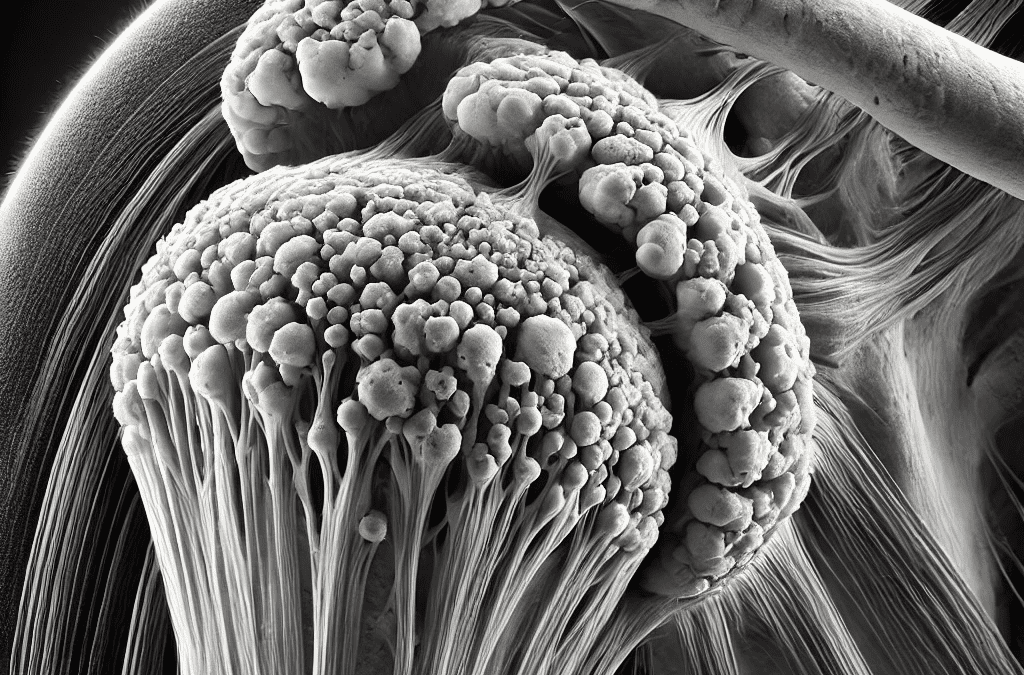
Calcific tendinitis of the rotator cuff (CTRC) is the accumulation of calcium phosphate within a tendon and is often chronic and recurrent. The majority of CTRC cases are located within 1–2 cm of the insertion of the supraspinatus, leading to a restricted range of motion in the shoulder and potentially causing severe pain (Maja et al., 2023). Interestingly, CTRC commonly occurs between the ages of 30 and 50 and is twice as likely to affect women (Kim et al., 2020). There are two proposed theories regarding the formation of CTRC: the degenerative theory and the reactive theory. The degenerative theory suggests that age-related changes in the tendon lead to a reduction in blood vessel distribution. This results in a hypoxic environment, causing necrosis and/or tendon tearing, which can subsequently lead to calcification (Kim et al., 2020). The reactive theory, on the other hand, describes three phases of calcification: the precalcific phase, the calcific phase, and the post-calcific phase. The precalcific phase involves the ‘transformation of the tendon into fibrocartilaginous tissue’ (Maja et al., 2023), facilitating calcium deposition. The calcific phase is when the actual deposition of calcium occurs, while the post-calcific phase involves the remodelling of the tendon around the calcium deposit. However, neither theory has been definitively proven (Maja et al., 2023). Secondary complications such as bursitis and synovitis are common in CTRC due to the chemical irritation caused by calcium deposits. Treatment for CTRC can be either conservative or surgical. Conservative management has a success rate of 30–80%. Non-steroidal anti-inflammatory drugs (NSAIDs) provide effective pain relief, and corticosteroid injections are beneficial during the resorptive phase. Barbotage has been shown to relieve pain in 70% of patients due to its decompression effects. Ultrasound therapy has also been found to improve quality of life and pain relief; however, it requires regular attendance over a six-week period. Extracorporeal shockwave therapy (ESWT) has the highest success rate for chronic calcific tendinitis and achieves results comparable to surgery (Kim et al., 2020). ESWT is effective in improving function and reducing pain, with greater efficacy when combined with physiotherapy (Maja et al., 2023). Therefore, conservative treatment should be prioritised and implemented for at least six months before considering surgical intervention (Maja et al., 2023). If you are experiencing symptoms of calcific tendinitis, it is important to seek professional medical advice. Booking an appointment with sports medicine doctor Dr David Porter (link to bio) can help you explore a range of treatment options tailored to your specific condition. Early intervention and expert guidance can significantly improve your recovery and long-term shoulder health.

Reference List

Kim, M.-S., Kim, I.-W., Lee, S. and Shin, S.-J. (2020). Diagnosis and treatment of calcific tendinitis of the shoulder. Clinics in Shoulder and Elbow, [online] 23(4), pp.203–209. doi:https://doi.org/10.5397/cise.2020.00318. Маја Manoleva, Erieta Nikolic Dimitrova, Koevska, V., Biljana Mitrevska, Marija Gocevska Gjerakaroska, Cvetanka Savevska, Biljana Kalchovska Ivanovska, Lidija Stojanoska Matjanoska, Gecevska, D., Jugova, T. and Liljana Malinovska Nikolovska (2023). Comparison of Immediate Effects of Extracorporeal Shockwave Therapy and Conventional Physical Therapy in Patients with Calcific Tendinitis of the Shoulder Rotator Cuff. Academic Medical Journal, 3(1), pp.99–109. doi:https://doi.org/10.53582/amj2331099m.
A New Approach to Managing Osteoarthritis: The Role of Stem Cell Therapy and Regenerative Medicine

A New Approach to Managing Osteoarthritis: The Role of Stem Cell Therapy and Regenerative Medicine

Understanding Osteoarthritis and Its Impact

Osteoarthritis (OA) is one of the most common joint conditions, affecting millions of people worldwide. It develops when the protective cartilage that cushions the joints gradually wears away, leading to pain, stiffness, and reduced mobility. Over time, the condition can significantly impact daily activities, making tasks such as walking, climbing stairs, or even getting out of bed challenging.

Traditional treatments for OA include pain relief medications, physiotherapy , steroid injections, and, in severe cases, joint replacement surgery. While these treatments help manage symptoms, they do not address the underlying cause, cartilage loss. This has led to growing interest in regenerative medicine, an emerging field focused on helping the body repair itself.

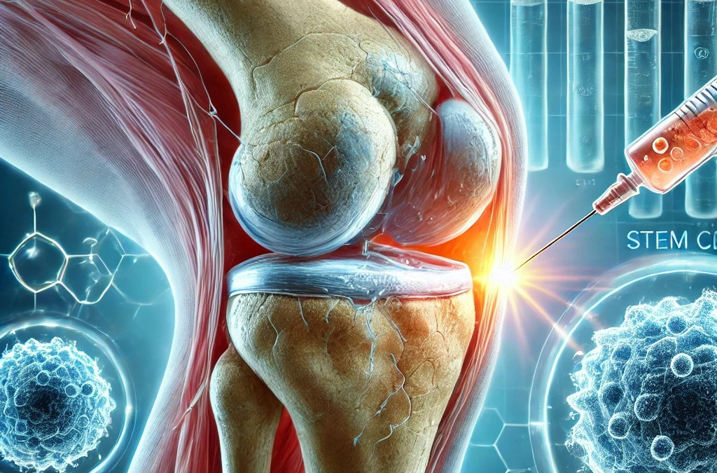
One area of regenerative medicine that has gained attention in recent years is stem cell therapy . Although this treatment is still undergoing research and development, early findings suggest it may offer a new approach to joint health by supporting tissue repair and reducing inflammation.

What Are Stem Cells?

Stem cells are special types of cells that have the potential to develop into different cell types in the body. This ability makes them important for healing and regeneration. The human body contains various types of stem cells, but mesenchymal stem cells (MSCs) are of particular interest when it comes to joint health.

MSCs can be obtained from several sources, including:

  • Bone marrow (from inside bones)
  • Adipose tissue (fat cells)
  • Umbilical cord tissue (from donated umbilical cords after birth)

Among these, umbilical cord-derived MSCs (UC-MSCs) have been widely studied for their potential role in joint repair.

How Can Stem Cells Help in Osteoarthritis?

While osteoarthritis is typically considered irreversible, ongoing research is exploring how stem cells might help slow its progression, reduce symptoms, and potentially support cartilage repair.

The Role of UC-MSCs in Joint Health

Umbilical cord-derived mesenchymal stem cells (UC-MSCs) are being studied for their ability to:

  • Reduce inflammation in the joint – Inflammation plays a key role in osteoarthritis and contributes to pain and stiffness.
  • Support cartilage maintenance – While research is ongoing, UC-MSCs are believed to release factors that encourage cartilage cells to survive and function.
  • Improve joint lubrication – UC-MSCs may help enhance the quality of synovial fluid, the natural lubricant in joints, improving movement.
  • Modulate the immune system – Osteoarthritis has been linked to immune system activity. UC-MSCs have the potential to balance immune responses in the joint.

Research into UC-MSCs is still ongoing, and while early results are promising, more studies are needed to determine their long-term effects and optimal use.

What Does the Latest Research Say?

Over the past few years, several studies have explored the potential of umbilical cord-derived stem cells in osteoarthritis treatment. Some notable findings include:

  1. Potential for Knee Cartilage Repair
    A review published in Medicine (2025) examined the use of UC-MSCs for knee osteoarthritis. The study found that these cells may contribute to reduced pain, improved function, and potential cartilage preservation. (Liao et al., 2025).
    📄 Read More: LWW Journal
  2. Stem Cell-Based Hydrogels for Cartilage Support
    A study in ACS Biomaterials Science (2025) investigated biodegradable hydrogels combined with UC-MSCs to enhance cartilage repair. The findings suggested improved joint function and reduced inflammation. (Ghosal et al., 2025).
    📄 Read More: ACS Publications
  3. Exosome Therapy for Osteoarthritis
    Scientists are also studying exosomes—tiny particles released by UC-MSCs – which may have anti-inflammatory and regenerative effects. A study in Advanced Functional Materials (2025) showed that exosomes from UC-MSCs supported cartilage repair and joint function in osteoarthritis models. (Lv et al., 2025).
    📄 Read More: Wiley Online Library
  4. UC-MSCs and Joint Injections
    Clinical trials published in Stem Cell Research & Therapy (2025) evaluated the safety and effects of UC-MSC injections for knee OA. The study noted improvements in pain, mobility, and joint function after six months. (Lei et al., 2025).
    📄 Read More: Springer

These studies represent a growing body of research exploring how stem cells may contribute to joint health and osteoarthritis management.

Important Considerations

While the idea of regenerative treatments for osteoarthritis is exciting, there are a few important points to keep in mind:

1. Research is Ongoing

Stem cell treatments, including those using umbilical cord-derived stem cells, are still being researched. Scientists continue to investigate their effectiveness, best application methods, and long-term safety.

2. Treatments Are Not Yet Widely Approved

While some countries have introduced regenerative medicine into clinical practice, regulatory authorities such as the UK’s MHRA (Medicines and Healthcare products Regulatory Agency) are still assessing these treatments. Opus, unlike many other clinics, is the UKs only licenced and regulated provider. It is essential to stay informed and seek advice from medical professionals before considering any new treatment options.

3. Every Patient is Different

Osteoarthritis can vary widely between individuals, meaning that treatment responses may differ. What works for one person may not work in the same way for another.

4. A Holistic Approach is Best

Stem cell therapy, if proven to be effective in the future, is unlikely to be a standalone cure for osteoarthritis. It is always best considered alongside other approaches such as:

  • Maintaining a healthy weight to reduce joint strain.
  • Engaging in regular, low-impact exercise (such as swimming or cycling).
  • Using joint-friendly supplements (such as glucosamine and omega-3 fatty acids).
  • Following a balanced diet to support joint health.


The Future of Regenerative Medicine

Research into umbilical cord-derived stem cells for osteoarthritis is advancing rapidly, with promising early results. Scientists are also exploring combination therapies, including:

  • Stem cells with growth factors to enhance repair.
  • 3D-printed cartilage scaffolds to create personalised joint implants.
  • Gene editing to enhance stem cell performance.

As research continues, the goal is to develop treatments that not only relieve symptoms but also support the long-term health of joints.

Conclusion

Osteoarthritis remains a challenging condition, but advances in stem cell research and regenerative medicine are opening up new possibilities. While umbilical cord-derived stem cells are still being studied, they hold promise as a potential tool for reducing inflammation, supporting joint function, and promoting cartilage health.

For now, education and staying informed about scientific advancements are key. As more research emerges, individuals seeking alternative approaches to joint care will have a better understanding of the options available.

If you are considering different ways to manage osteoarthritis, always consult a healthcare professional for personalised advice.


References

  1. Liao, Z.K. et al. (2025). Clinical research progress of umbilical cord blood mesenchymal stem cells in knee articular cartilage repair. Medicine.
    📄 Read More
  2. Lei, J. et al. (2025). Therapeutic efficacy of intra-articular injection of UC-MSCs in knee OA. Stem Cell Research & Therapy.
    📄 Read More


Osteoarthritis