Shoulder Archives - Opus Biological
Calcific Tendinitis of the Rotator Cuff (CTRC)

Calcific Tendinitis of the Rotator Cuff (CTRC)

Calcific tendinitis of the rotator cuff (CTRC) is the accumulation of calcium phosphate within a tendon and is often chronic and recurrent. The majority of CTRC cases are located within 1–2 cm of the insertion of the supraspinatus, leading to a restricted range of motion in the shoulder and potentially causing severe pain (Maja et al., 2023). Interestingly, CTRC commonly occurs between the ages of 30 and 50 and is twice as likely to affect women (Kim et al., 2020). There are two proposed theories regarding the formation of CTRC: the degenerative theory and the reactive theory. The degenerative theory suggests that age-related changes in the tendon lead to a reduction in blood vessel distribution. This results in a hypoxic environment, causing necrosis and/or tendon tearing, which can subsequently lead to calcification (Kim et al., 2020). The reactive theory, on the other hand, describes three phases of calcification: the precalcific phase, the calcific phase, and the post-calcific phase. The precalcific phase involves the ‘transformation of the tendon into fibrocartilaginous tissue’ (Maja et al., 2023), facilitating calcium deposition. The calcific phase is when the actual deposition of calcium occurs, while the post-calcific phase involves the remodelling of the tendon around the calcium deposit. However, neither theory has been definitively proven (Maja et al., 2023). Secondary complications such as bursitis and synovitis are common in CTRC due to the chemical irritation caused by calcium deposits. Treatment for CTRC can be either conservative or surgical. Conservative management has a success rate of 30–80%. Non-steroidal anti-inflammatory drugs (NSAIDs) provide effective pain relief, and corticosteroid injections are beneficial during the resorptive phase. Barbotage has been shown to relieve pain in 70% of patients due to its decompression effects. Ultrasound therapy has also been found to improve quality of life and pain relief; however, it requires regular attendance over a six-week period. Extracorporeal shockwave therapy (ESWT) has the highest success rate for chronic calcific tendinitis and achieves results comparable to surgery (Kim et al., 2020). ESWT is effective in improving function and reducing pain, with greater efficacy when combined with physiotherapy (Maja et al., 2023). Therefore, conservative treatment should be prioritised and implemented for at least six months before considering surgical intervention (Maja et al., 2023). If you are experiencing symptoms of calcific tendinitis, it is important to seek professional medical advice. Booking an appointment with sports medicine doctor Dr David Porter can help you explore a range of treatment options tailored to your specific condition. Early intervention and expert guidance can significantly improve your recovery and long-term shoulder health.

Reference List

Kim, M.-S., Kim, I.-W., Lee, S. and Shin, S.-J. (2020). Diagnosis and treatment of calcific tendinitis of the shoulder. Clinics in Shoulder and Elbow, [online] 23(4), pp.203–209. doi:https://doi.org/10.5397/cise.2020.00318. Маја Manoleva, Erieta Nikolic Dimitrova, Koevska, V., Biljana Mitrevska, Marija Gocevska Gjerakaroska, Cvetanka Savevska, Biljana Kalchovska Ivanovska, Lidija Stojanoska Matjanoska, Gecevska, D., Jugova, T. and Liljana Malinovska Nikolovska (2023). Comparison of Immediate Effects of Extracorporeal Shockwave Therapy and Conventional Physical Therapy in Patients with Calcific Tendinitis of the Shoulder Rotator Cuff. Academic Medical Journal, 3(1), pp.99–109. doi:https://doi.org/10.53582/amj2331099m.
Calcific Tendinitis of the Rotator Cuff (CTRC)

Calcific Tendinits of the rotator cuff

Calcific Tendinits of the rotator cuff (CTRC) is the accumulation of calcium phosphate within a tendon and is often chronic and recurrent. Majority of CTRC are located within 1-2cm from the insertion of the supraspinatus causing limited range of motion in the shoulder and can cause severe pain (Maja et. al, 2023). Interestingly, CTRC commonly occurs during the age of 30-50 years and twice as likely to occur in women (Kim et. al, 2020).

There are two proposed theories on the formation of CTRC: degenerative and reactive. Degenerative theory suggests changes in the tendon accumulate with age which causes a decrease in distribution of blood vessels. This leads to a hypoxic environment causing necrosis and/or tearing in the tendon which then can develop into calcification (Kim et. al, 2020). The reactive theory suggests that there are 3 phases of calcification: precalcifying phase, calcifying phase and post calcifying phase. Precalcifying phase is the ‘transformation of the tendon into fibrocartilaginous tissue’ (Maja et. al, 2023) which allows the calcium to be deposited more easily. The calcifying phase is the actual deposition of calcium, and the post calcifying is the remodeling of the tendon around the calcium deposit. However, no theory has been proven (Maja et. al, 2023). Secondary complications such as bursitis and synovitis are common with CTRC due to the chemical irritation caused by calcium deposits.

Treatment for CTRC can either be conservative management or surgical. There is a 30-80% success rate of conservative management. NSAIDs are effective for pain relief as well as a steroid injection during the resorptive phase. Barbotage has been shown to provide pain relief in 70% of patients due to its decompression effects. Ultrasound therapy has also been shown to improve quality of life and pain relief, however, requires regular attendance over a 6 week period. ESWT (extracorporeal shockwave therapy) has the highest success rate for chronic calcific tendinitis and has a similar success rate to surgery (Kim et. al, 2020). ESWT is effective in improving function and reducing pain and is more effective when combined with physiotherapy (Maja et. al, 2023). Therefore, conservative treatment should be prioritized and conducted for a minimum of 6 months before considering surgical treatment (Maja et. al, 2023).

Reference List

Kim, M.-S., Kim, I.-W., Lee, S. and Shin, S.-J. (2020). Diagnosis and treatment of calcific tendinitis of the shoulder. Clinics in Shoulder and Elbow, [online] 23(4), pp.203–209. doi:https://doi.org/10.5397/cise.2020.00318.

Маја Manoleva, Erieta Nikolic Dimitrova, Koevska, V., Biljana Mitrevska, Marija Gocevska Gjerakaroska, Cvetanka Savevska, Biljana Kalchovska Ivanovska, Lidija Stojanoska Matjanoska, Gecevska, D., Jugova, T. and Liljana Malinovska Nikolovska (2023). COMPARISON OF IMMEDIATE EFFECTS OF EXTRACORPOREAL SHOCKWAVE THERAPY AND CONVENTIONAL PHYSICAL THERAPY IN PATIENTS WITH CALCIFIC TENDINITIS OF THE SHOULDER ROTATOR CUFF. Academic Medical Journal, 3(1), pp.99–109. doi:https://doi.org/10.53582/amj2331099m.

Calcific Tendinitis of the Rotator Cuff (CTRC)
Common shoulder injuries amongst athletes and how we treat them

Common shoulder injuries amongst athletes and how we treat them

Black and white image of an athlete lifting weights
Photo by Quino Al on Unsplash

Acromioclavicular joint injury

The acromioclavicular (AC) joint is the point at which two bones, the collarbone (clavicle) and part of the shoulder blade (scapula), meet. An injury to this joint is characterised by shoulder separation and can be defined as one of six types. The severity of the injury also impacts the treatment approach.
1. Type I. The AC ligament is slightly torn; however, there is no significant damage to the coracoclavicular ligaments. Treatment often centres around rest and recovery, with the joint being iced and protected, often with an arm swing. Following this, range-of-motion exercises are recommended as soon as they can be tolerated. 2. Type II. The AC ligament is completely torn; however, there is little to no tear to the coracoclavicular ligaments. Initial treatment involves rest, ice, pain medication, and up to a week of shoulder immobilisation in a sling. Range-of-motion exercises are recommended alongside strengthening exercises.
3. Type III. The AC and coracoclavicular ligaments are completely torn, resulting in the collarbone separating from the scapula. Although treatment for this injury can be conducted non-surgically, as with type I and II injuries, the duration of recovery is significantly longer, with a sling being required for up to a month. 4. Type IV, V, VI. Treatment of these injuries almost exclusively involves surgical interventions, with specialists required to reduce the risk of long-term complications. In these cases, regenerative medicine may also be considered.

Rotator cuff tendonitis

Rotator cuff tendonitis is characterised by pain and swelling of the cuff tendons and the surrounding bursa (a soft fluid filled sack that cushions the joint). This injury, unlike an injury to the AC joint, does not occur all at once. Usually, rotator cuff tendonitis transpires over a substantial period of time following repeated irritation to the area. Although it can affect anyone, this injury most commonly presents in people with loose joints, abnormal bone anatomy in the shoulder, and those who do repetitive heavy lifting, such as weightlifters and powerlifters.
Treatment of rotator cuff tendonitis is predominantly non-surgical and involves plenty of ice, NSAIDS for pain management, including ibuprofen, and steroids. In the instance that surgery is required, the most common procedure is an acromioplasty. However, recent advances in regenerative medicine have permitted the accelerated and enhanced recovery of these injuries using mesenchymal stem cell therapy.

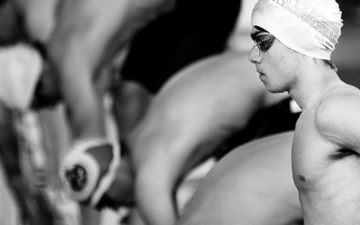
Beyond these individual conditions described, there are also activity-specific injuries, including thrower’s shoulder, swimmer’s shoulder, and rugby shoulder. At Opus, we ensure that your injury is fully assessed so that you receive the best possible treatment that is tailored to your needs. Get in touch to discuss your recovery with one of our world renowned specialists.