The impact of fatigue and why optimal recovery is so important.
Optimal recovery from training and competition is most definitely a subject that we are seeing more frequently discussed and addressed in elite sport and the sports medicine world. Away from sporting competition, we can also inform and educate individuals on how to ensure their recovery is as good as it can be to enable them to be on the top of their game in their day to day life whether that be at home or at work. With useful data collecting tools such as; the Whoop, Oura ring and Apple watch, it is becoming much easier for people to keep an eye on their vital metrics and determine when rest is needed or when is a good time to place their body under higher strain through training, competition or a stressful life event.
Here at Opus, we often have the ‘optimal recovery’ discussion with our patients. It tends to be part of a greater conversation of how we assist in managing that individual’s training load and or managing their current state of physical and mental health. By doing this we can reduce the risk of injury and illness and aid in the patient performing to the best of their ability in whatever task it may be that matters to them.
So what is optimal recovery? The fact is, it is not just any one thing, but instead a combination of a number of components that contribute to restoring a person’s physical, and mental well-being after a period of physical or mental exertion. By ‘optimising’ as many of these components as possible, we can in turn reduce the potential risk of both illness and injury.
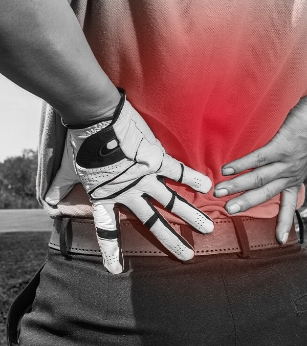
Two specific musculoskeletal injuries linked to fatigue which have been researched in depth are hamstring and ACL injuries. One study by Mclean and Samorezov (2009) (2), reported that the inhibitory action of the fatigued muscles in landing activities, increases the possibility of ineffective perception, decision, and movement execution strategies therefore increasing risk of injury to the ACL. Small et. Al (2009)(3) focused their investigation on the risk of hamstring injury at the ends of each half of a football match. Their findings suggest that due to decreased flexibility of the hamstring muscle group during fatigued sprinting, with a reduced maximum combined hip flexion and knee extension angle observed during the late swing phase of the sprinting cycle, the strain on the hamstring muscle group is increased in this fatigued state and the risk of hamstring injury is amplified.
Jones et. Al (2016)(1) looked more into the impact of training load on the risk of illness due to fatigue. They found that during periods of training load intensification, or where there was a greater accumulation of training load, the risk of acquiring illness increases.
So what are these key components of optimal recovery and how can they impact an individual? Some of those we discuss regularly with our patents at Opus are:
- Getting sufficient rest and good quality sleep are fundamental for recovery. Sleep is when the body repairs tissues, consolidates memories, and regulates the hormones which are crucial for recovery and overall health.
- Being hot on your nutrition. A good diet focused around the right food types, supports the body’s recovery processes and replenishes energy stores, repairs tissues, and supports immune function.
- Maintaining proper hydration levels. Water is crucial for transporting nutrients, regulating body temperature, and facilitating various biochemical processes in the body.
- Opting for ‘Active Recovery’ when necessary. A pool session, stretch and mobility routine or flush on the watt bike, can enhance circulation, reduce muscle stiffness, and promote recovery by aiding in the removal of metabolic waste products from muscles.
- Managing stress. Using techniques such as meditation or deep breathing exercises, can help reduce stress levels and promote recovery. Alternatively if that’s not for you, surrounding yourself with friends and family and taking part in activities that you find fun and relaxing can be of great help.
So in short, what we do away from the more strenuous or testing activities in our day to day life, is vital in ensuring our performance is at its best. Optimal recovery leads to reduced risk of injury and illness and in turn, optimal performance.
Reference List
- Jones, C.M., Griffiths, P.C. and Mellalieu, S.D. (2016). Training Load and Fatigue Marker Associations with Injury and Illness: A Systematic Review of Longitudinal Studies. Sports Medicine, 47(5), pp.943–974. doi:https://doi.org/10.1007/s40279-016-0619-5.
- MCLEAN, S.G. and SAMOREZOV, J.E. (2009). Fatigue-Induced ACL Injury Risk Stems from a Degradation in Central Control. Medicine & Science in Sports & Exercise, 41(8), pp.1661–1672. doi:https://doi.org/10.1249/mss.0b013e31819ca07b.
- Small, K., McNaughton, L.R., Greig, M., Lohkamp, M. and Lovell, R. (2009). Soccer Fatigue, Sprinting and Hamstring Injury Risk. International Journal of Sports Medicine, 30(08), pp.573–578. doi:https://doi.org/10.1055/s-0029-1202822.
")