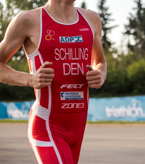
opusbiological, Author at opusbiological.com - Page 7 of 9
Plantar fasciitis: A common injury causing global heel pain

Plantar fasciitis: A common injury causing global heel pain

Photo by Tonnam Vongsamang on Unsplash

Plantar fasciitis holds the title as one of the most prevalent causes of heel pain across the globe. It is characterised by inflammation and causes severe stabbing pain that usually occurs with your first steps of the day. Movement does, however, result in a decrease in pain throughout the day. Although the underlying causes of this injury are poorly comprehended, it is more common in runners and older adults. Current statistics suggest that the prevalence of plantar heel pain in the UK population is 9.6%, with the population prevalence of disabling plantar heel pain at 7.9%.

The plantar fascia is a band of tissue that connects the heel bone to the base of the toes, supporting the foot’s arch and absorbing shock with every step we take. However, prolonged tension and stress on this band of tissue can cause small tears. Alone, they do not cause significant injury, but repeated stretching and tearing of the fascia can cause irritation and inflammation, characteristic of plantar fasciitis. 

Treatment of plantar fasciitis is varied and depends on the severity of pain experienced. There are remedies that can be adopted at home, or more specialist-focused approaches, including stem cell injections.

  • Physical therapy is the most commonly adopted treatment option for plantar fasciitis; however, it does not fix the root cause of the problem, and many people experience recurrence of the pain further down the line. Exercises such as heel raise, floor sitting ankle inversion with resistance, seated toe towel scrunches, and plantar fascia stretches can all assist in easing plantar heel pain.
  • Steroid injections, specifically cortisone injections, offer short-term pain relief of a few months. However, the risks associated with this method, including skin thinning, often deter people from taking this approach.
  • Plantar fasciotomy is a surgical intervention that detaches the fascia from the heel bone in order to relieve tension and provide a long-term pain relief option. Despite this, only about 5% of those with plantar fasciitis choose this approach as it is only suggested if you experience persistent heel pain for greater than six months.
  • Stem cell injections and platelet rich plasma form the regenerative approach to plantar fasciitis treatment. The goal of these interventions is to expedite your recovery journey and provide a therapeutic option with more long-lasting results. You can find more information on how these regenerative approaches work in our previous posts.

At Opus, we offer both platelet rich plasma therapy and stem cell therapy to combat your plantar heel pain. Get in touch to speak to one of our experts and begin your journey to pain free movement.

Lower Back Pain and Pelvic Floor Dysfunction

Lower Back Pain and Pelvic Floor Dysfunction

Lower back pain (LBP) is the most prevalent musculoskeletal injury, as approximately 80% of the population will experience LBP in their lifetime (Arab et. al 2010). Transverse abdominus activation is often prescribed for LBP however pelvic floor muscle (PFM) is not always incorporated into treatment. PFM aids in supporting the abdominopelvic organs and there is an abundance of research on the role of the PFM in urinary and fecal incontinence. Nevertheless, it is important to remember the PFM role in lumbar and pelvic stability and intra-abdominal pressure (Mohseni-Bandepi et. al 2011). Research illustrates that the combination of pelvic floor exercise alongside routine treatment can provide significant pain relief in LBP compared to routine treatment alone (Bi et. al, 2013), demonstrating the importance of PFM exercises.

The link between LBP and PFM helps to provide insight into the correlation between LBP and pelvic floor dysfunction (PFD). PFD is an umbrella term to describe weakness, poor endurance and hypertonicity of the PFM which can impact incontinence, prolapses and pelvic pain. Dufour et al. found that 95% of women with LBP had PFD, ‘71% of the participants had pelvic floor muscle tenderness, 66% had pelvic floor weakness and 41% were found to have a pelvic organ prolapse’(Dufour et. al 2018), further highlighting the importance of incorporating PFM in treatment. Arab et al. also examined PFD in women with and without LBP. The results show PFD in participants with LBP compared to those without. This is valuable for health care participants when assessment and treating LBP (Arab et. al 2010). It is also important to recognize PFD in pregnancy-related lower back pain (PLBP). Pool-Goudzwaard et. al discovered ‘52% of PLBP, significantly more than in the healthy control group’ (Pool-Goudzwaard et. al,2005) and promote addressing both LBP and PFD during pregnancy.

There are a variety of methods to help activate, strengthen and relax the pelvic floor in order to aid recovery in PFD.  Kegels or reverse Kegels are often prescribed to promote awareness and strengthen PFM (Torgenu et. Al 2021). Transverse abdominus contraction has also been shown to help activate and strengthen PFM. Sapsford et al. found ‘that abdominals contract in response to a pelvic floor contraction command and that the pelvic floor contracts in response to both a “hollowing” and “bracing” abdominal command’(Mohseni-Bandepi et. al 2011), therefore proving the PFM can be activated by engaging the abdominals. Hypopressive exercises (HE) have also shown to aid recovery from PFD. HEs lowers the intra-abdominal pressure which enables an involuntary contraction of the PFM and transverse abdominus. Navarro-Brazález et. al suggests the combination of HEs and pelvic floor muscle training improves PFM strength and quality of life for patients with PFD (Navarro-Brazález et. al 2020).

Reference List


  • Arab, A.M., Behbahani, R.B., Lorestani, L. and Azari, A. (2010). Assessment of pelvic floor muscle function in women with and without low back pain using transabdominal ultrasound. Manual Therapy, 15(3), pp.235–239. doi:https://doi.org/10.1016/j.math.2009.12.005.

  • Bi, X., Zhao, J., Zhao, L., Liu, Z., Zhang, J., Sun, D., Song, L. and Xia, Y. (2013). Pelvic floor muscle exercise for chronic low back pain. Journal of International Medical Research, 41(1), pp.146–152. doi:https://doi.org/10.1177/0300060513475383.

  • Dufour, S., Vandyken, B., Forget, M.-J. and Vandyken, C. (2018). Association between lumbopelvic pain and pelvic floor dysfunction in women: A cross sectional study. Musculoskeletal Science and Practice, 34, pp.47–53. doi:https://doi.org/10.1016/j.msksp.2017.12.001.

  • Mohseni-Bandpei, M.A., Rahmani, N., Behtash, H. and Karimloo, M. (2011). The effect of pelvic floor muscle exercise on women with chronic non-specific low back pain. Journal of Bodywork and Movement Therapies, 15(1), pp.75–81. doi:https://doi.org/10.1016/j.jbmt.2009.12.001.

  • Navarro-Brazález, B., Prieto-Gómez, V., Prieto-Merino, D., Sánchez-Sánchez, B., McLean, L. and Torres-Lacomba, M. (2020). Effectiveness of Hypopressive Exercises in Women with Pelvic Floor Dysfunction: A Randomised Controlled Trial. Journal of Clinical Medicine, 9(4), p.1149. doi:https://doi.org/10.3390/jcm9041149.

  • Pool-Goudzwaard, A.L., Slieker ten Hove, M.C.P.H., Vierhout, M.E., Mulder, Paul.H., Pool, J.J.M., Snijders, C.J. and Stoeckart, R. (2005). Relations between pregnancy-related low back pain, pelvic floor activity and pelvic floor dysfunction. International Urogynecology Journal, [online] 16(6), pp.468–474. doi:https://doi.org/10.1007/s00192-005-1292-7.

  • Torgbenu, E.L., Aimakhu, C.O. and Morhe, E.K.S. (2020). Effect of Kegel Exercises on Pelvic Floor Muscle Disorders in Prenatal and Postnatal Women – A Literature Review. Current Women’s Health Reviews, 16. doi:https://doi.org/10.2174/1573404816999200930161059.

Pain down there? Common causes and treatment of groin pain

Pain down there? Common causes and treatment of groin pain

Photo by Victoire Joncheray on Unsplash

In the vast majority of cases, groin pain results from a muscle strain or tear and usually resolves on its own. Athletes disproportionately experience groin pain due to their activity levels, with statistics suggesting prevalence rates between 0.5% and 28% amongst football players. But what actually causes groin pain in the first place?

There are several unique causes of groin pain; however, the most common cause is a muscle strain, also known as a pull groin muscle. This occurs when the hip joint is involved in an acquired movement that causes a stretch or tears to the inner thigh muscles. The pain experienced is usually sharp and sudden, with the source of it often apparent. Inner thigh muscle spasms and leg weakness may also occur.

Beyond muscle strain, the following are also common causes of groin pain:

  • Inguinal hernia. A condition where the fatty tissue of the intestine protrudes through the abdominal wall.
  • Kidney stone. A small clump of minerals will form inside the kidney, causing waves of pain as it passes down through the urinary tract.
  • Hip osteoarthritis arises when the smooth hip joint is worn down, causing leg movements to become painful and stiff.

Treating groin pain depends on what started it in the first place. In most instances, you may not need medical intervention; however, if the pain persists and impacts your daily activities, then get in touch.

When determining the best route of treatment, you will be asked when the pain started, what activities exacerbate the pain, and if you have experienced any accompanying symptoms. A full physical examination will then be performed, with imaging often being used as well.

There are five treatment approaches that may be taken, depending on the severity of the pain and your lifestyle:

    1. Rest and recuperation. Although it seems repetitive with sports injuries, rest and ice are your best friends. Limiting your activities as much as possible for a short period of time can significantly speed up the healing process.
    2. This approach solely offers short-term pain relief and has no significant impact on the healing process.
    3. Physical therapy. Depending on the cause of the groin pain, you may be referred for physical therapy. For example, if your groin pain results from hip-related causes, such as hip osteoarthritis, then long-term physical therapy may be beneficial in managing flare ups. Moreover, certain exercises can help improve your range of motion and flexibility, further preventing further injuries.
    4. This intervention is not always necessary and is only applicable in some serious conditions, including hip joint infections.
    5. Regenerative medicine. Platelet rich plasma (PRP) is a novel treatment that has had great success in treating arthritic conditions, including hip osteoarthritis. We go into more detail on the process of PRP here. The benefits of PRP on hip function and pain have been shown to be superior to other options given the long-term duration of its effects.

    At Opus, we pride ourselves on offering a range of treatments to our patients to ensure that the most appropriate route of recovery is selected. If you are experiencing persistent groin pain, we can provide you with the traditional routes of treatment but also the regenerative options, including PRP. Get in touch today to discuss the best option to get you back on your feet.

    Lower Back Pain and Pelvic Floor Dysfunction

    The impact of fatigue and why optimal recovery is so important.

    Optimal recovery from training and competition is most definitely a subject that we are seeing more frequently discussed and addressed in elite sport and the sports medicine world. Away from sporting competition, we can also inform and educate individuals on how to ensure their recovery is as good as it can be to enable them to be on the top of their game in their day to day life whether that be at home or at work. With useful data collecting tools such as; the Whoop, Oura ring and Apple watch, it is becoming much easier for people to keep an eye on their vital metrics and determine when rest is needed or when is a good time to place their body under higher strain through training, competition or a stressful life event.

    Here at Opus, we often have the ‘optimal recovery’ discussion with our patients. It tends to be part of a greater conversation of how we assist in managing that individual’s training load and or managing their current state of physical and mental health. By doing this we can reduce the risk of injury and illness and aid in the patient performing to the best of their ability in whatever task it may be that matters to them.

    So what is optimal recovery? The fact is, it is not just any one thing, but instead a combination of a number of components that contribute to restoring a person’s physical, and mental well-being after a period of physical or mental exertion. By ‘optimising’ as many of these components as possible, we can in turn reduce the potential risk of both illness and injury.

    Two specific musculoskeletal injuries linked to fatigue which have been researched in depth are hamstring and ACL injuries. One study by Mclean and Samorezov (2009) (2), reported that the inhibitory action of the fatigued muscles in landing activities,  increases the possibility of ineffective perception, decision, and movement execution strategies therefore increasing risk of injury to the ACL. Small et. Al (2009)(3) focused their investigation on the risk of hamstring injury at the ends of each half of a football match. Their findings suggest that due to decreased flexibility of the hamstring muscle group during fatigued sprinting, with a reduced maximum combined hip flexion and knee extension angle observed during the late swing phase of the sprinting cycle, the strain on the hamstring muscle group is increased in this fatigued state and the risk of hamstring injury is amplified.

    Jones et. Al (2016)(1) looked more into the impact of training load on the risk of illness due to fatigue. They found that during periods of training load intensification, or where there was a greater accumulation of training load, the risk of acquiring illness increases.

    So what are these key components of optimal recovery and how can they impact an individual? Some of those we discuss regularly with our patents at Opus are:

    • Getting sufficient rest and good quality sleep are fundamental for recovery. Sleep is when the body repairs tissues, consolidates memories, and regulates the hormones which are crucial for recovery and overall health.
    • Being hot on your nutrition. A good diet focused around the right food types, supports the body’s recovery processes and replenishes energy stores, repairs tissues, and supports immune function.
    • Maintaining proper hydration levels. Water is crucial for transporting nutrients, regulating body temperature, and facilitating various biochemical processes in the body.
    • Opting for ‘Active Recovery’ when necessary. A pool session, stretch and mobility routine or flush on the watt bike, can enhance circulation, reduce muscle stiffness, and promote recovery by aiding in the removal of metabolic waste products from muscles.
    • Managing stress. Using techniques such as meditation or deep breathing exercises, can help reduce stress levels and promote recovery. Alternatively if that’s not for you, surrounding yourself with friends and family and taking part in activities that you find fun and relaxing can be of great help.

    So in short, what we do away from the more strenuous or testing activities in our day to day life, is vital in ensuring our performance is at its best. Optimal recovery leads to reduced risk of injury and illness and in turn, optimal performance.

    Reference List


    • Jones, C.M., Griffiths, P.C. and Mellalieu, S.D. (2016). Training Load and Fatigue Marker Associations with Injury and Illness: A Systematic Review of Longitudinal Studies. Sports Medicine, 47(5), pp.943–974. doi:https://doi.org/10.1007/s40279-016-0619-5.
    • MCLEAN, S.G. and SAMOREZOV, J.E. (2009). Fatigue-Induced ACL Injury Risk Stems from a Degradation in Central Control. Medicine & Science in Sports & Exercise, 41(8), pp.1661–1672. doi:https://doi.org/10.1249/mss.0b013e31819ca07b.
    • Small, K., McNaughton, L.R., Greig, M., Lohkamp, M. and Lovell, R. (2009). Soccer Fatigue, Sprinting and Hamstring Injury Risk. International Journal of Sports Medicine, 30(08), pp.573–578. doi:https://doi.org/10.1055/s-0029-1202822.

    Debunking myths about PRP

    Debunking myths about PRP

    Photo by Kindel Media on Pexels
    PRP is a minimally invasive outpatient procedure and can reduce pain, improve function and help you return to your normal activities or sporting pursuits. This novel approach, despite its success in clinical studies, is still surrounded by several myths. So, let’s debunk them.

    1. PRP therapy requires surgery.

    The plasma, the liquid where the platelets reside, is drawn from your body before being placed in a machine that separates the PRP from the rest of the blood. This PRP is then injected into the area of your body being treated. Once the platelets are in the target area, they will break down, releasing growth factors, important proteins that stimulate cell growth, differentiation, survival, inflammation, and tissue repair; collectively triggering your body’s healing process.
    As you can see, PRP therapy does not require surgery, solely injections. In most instances, real-time imaging will also be used to guide us and ensure that the PRP is injected directly at the site of your injured tissues.

    2. PRP is only good for pain relief.

    Although PRP is generally used in patients with chronic pain conditions, there are so many additional benefits that this treatment will present beyond pain relief. This includes reduced inflammation, the recruitment of stem cells to the area triggering new tissue production, stimulation of blood vessel growth, and the degeneration prevention of healthy tissues. These benefits combine to speed up tissue regeneration and healing, relieving your pain. This is especially beneficial for athletes who are looking to get back on their feet as soon as possible.

    3. PRP is a highly risky procedure.

    Given that PRP therapy relies solely on injections, the procedure itself is deemed exceptionally safe. For starters, the plasma injected is autologous, meaning that it is made for your own blood, and you do not need to worry about potential negative reactions or side effects. What’s more, PRP therapy has been used for several years now, with no major complications being reported.

    One study, in particular, investigated the clinical application and safety of PRP therapy for patients with patellar tendinopathy, a condition that causes chronic back pain. One of the key points the researchers noted, besides the remarkable improvement in back pain, was the safety of PRP and that no complications or side effects were seen across all 15 clinical studies included.

    Considering PRP therapy but don’t know where to start? Or have more questions that you need answering? Get in touch with our team to discuss your options and how we can help relieve your pain.